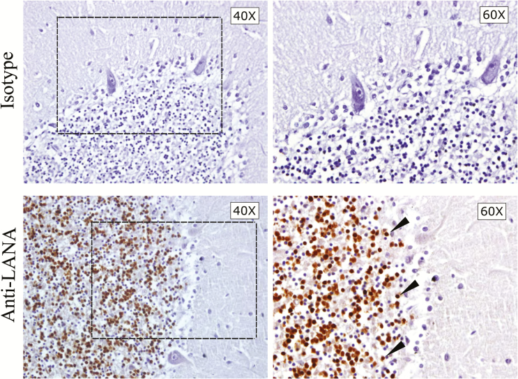
Figure 2.
Immunohistochemical staining for latency-associated nuclear antigen (LANA) on cerebellum (Cer) sections from subject 92. Sections within 30 µm of one another were stained with isotype control sera or anti-LANA. The different magnifications are of the same cerebellar folium in near-adjacent sections. The box indicates the selected area of the granular layer magnified in the right panel. LANA-positive cells appeared as brown punctate staining, and representative cells are designated by arrowheads.

