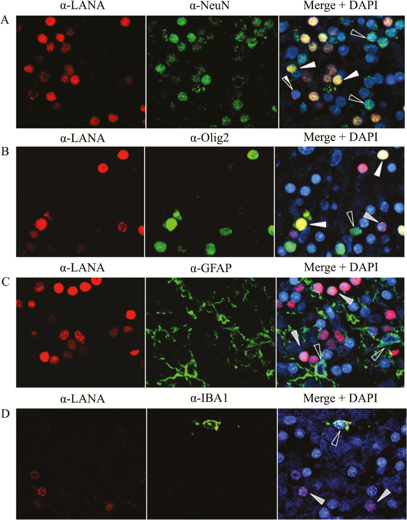
Figure 3.
Identification of Kaposi’s sarcoma–associated herpesvirus (KSHV)–infected cell types by immunofluorescence. Formalin-fixed, paraffin-embedded blocks of cerebellum tissue from subject 92 were divided into 5.0-µm sections and subjected to dual-label immunofluorescence, using mouse monoclonal anti-LANA (α-LANA) to identify cells expressing latent KSHV antigen, and cell lineage-specific antibodies directed against antigens for neurons (α-NeuN), oligodendrocytes (α-Olig2), astrocytes (α-GFAP), or activated macrophage/microglia (α-IBA1). White arrows denote infected cells costained with the lineage marker, gray arrows indicate infected cells of alternative lineage, and open arrows indicate lineage-identified uninfected cells.

