Figure 2.
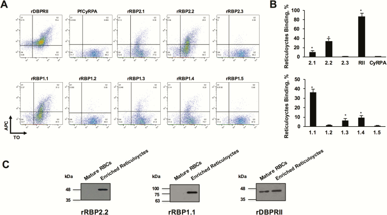
Flow cytometry–based reticulocyte-binding activity of Plasmodium vivax reticulocyte-binding protein (PvRBP) 2c and PvRBP1a recombinant fragments. (A) Flow cytometry dot plots showing the binding of rRBP2.1, rRBP2.2, rRBP2.3, rDBPRII, and PfCyRPA (top) and rRBP1.1, rRBP1.2, rRBP1.3, rRBP1.4, rRBP1.5 (bottom) with enriched reticulocytes (thiazole orange [TO] positive). Reticulocyte binding was detected using the antibodies raised against the respective recombinant proteins followed by a secondary allophycocyanin (APC)–conjugated monoclonal antibody. Of the 3 PvRBP2c proteins, reticulocyte binding was observed for rRBP2.2 and rRBP2.1, with no binding observed for rRBP2.3. Of the 5 PvRBP1a protein constructs, reticulocyte binding was observed for rRBP1.1, rRBP1.3, and rRBP1.4, with no binding reported for rRBP1.2 and rRBP1.5. Binding of rDBPRII and PfCyRPA was analyzed as a positive and negative controls, respectively. B, Bar charts depicting the percentage reticulocyte binding of rRBP2.1, rRBP2.2, rRBP2.3, rDBPRII, PfCyRPA (top) and rRBP1.1, rRBP1.2, rRBP1.3, rRBP1.4, and rRBP1.5 (bottom). Error bars represent standard errors of the mean for 3 independent repeats. *P < .001. C, Binding of recombinant rRBP2.2 and rRBP1.1 was detected only with enriched reticulocytes and not with normal erythrocytes (red blood cells [RBCs]. rDBPRII bound with the same set of normal erythrocytes and enriched reticulocytes. Reticulocytes were enriched by magnetic sorting. Abbreviations: PfCyRPA, Plasmodium falciparum cysteine-rich protective antigen; rDBPRII, recombinant Duffy binding protein region II; rRBP, recombinant RBP.

