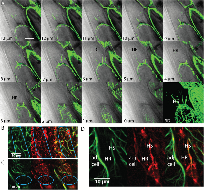
Fig. 7.
ER and actin filaments interaction during plasmolysis in Arabidopsis. A) Actin formation at different focal planes (lower left) of a plasmolyzed hypocotyl cell with a 3D reconstruction showing several Hechtian strands, one of which is labeled HS. Regions containing the Hechtian reticulum as identified with DIC are shown as HR. B–D) Confocal images of hypocotyl cells showing the separate green channel with actin filaments labeled with YFP-ABD2, the red channel showing the ER labeled with mCherry-HDEL, and a merged image. B) ER tracks on actin within the protoplast during plasmolysis. The outline of the protoplast is in cyan. C) Actin doesn’t show up in the Hechtian reticulum outlined in cyan. D) Actin forms a big Hechtian strand within the periplasmic region, labeled HS, but is excluded in the fine network of the Hechtian reticulum, labelled HR. Adj. cell, adjacent cell. Scale bar, 10 μm.

