Figure 3.
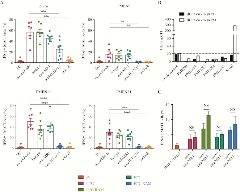
Mucosal-associated invariant T (MAIT) cell activation by pneumococci in presence of monocytes is not MR1-dependent. A, Paraformaldehyde-fixed Pneumococcal Molecular Epidemiological Network (PMEN) reference strains or Escherichia coli were cultured with peripheral blood mononuclear cells and THP1 cells in the presence of anti-MR1 blocking, anti–interleukin 12 (IL-12), and anti–interleukin 18 (IL-18) blocking antibodies. Interferon γ (IFN-γ) expression from MAIT cells is shown. ****P < .0001, ***P < .001, and **P < .01, by repeated measures 1-way analysis of variance (ANOVA) with the Dunnett multiple comparisons test, compared with the no antibody control (n = 7). B, Jurkat cells expressing the MAIT cell T-cell receptor (TCR; white bars) or control cells not expressing the MAIT cell TCR (black bars) were cultured with THP1 cells overnight with the indicated PMEN strains or E. coli as a positive control. Activation was measured as the geometric mean fluorescence intensity (gMFI) of CD69 expressed by Jurkat cells. The dotted line indicates value for the sterile control (SC). Data are representative of 3 independent experiments. C, The PMEN34 strain was grown overnight at 36°C or 40°C in Todd Hewitt broth with 0.5% yeast extract (THB-Y) and either cultured in THB-Y for the last 4 hours or transferred to riboflavin-free medium (RAM). The bacteria were then fixed and cultured with PBMCs and THP1 cells overnight. The frequency of IFN-γ–expressing MAIT cells is shown in the presence or absence of anti-MR1 blocking antibody. NS, nonsignificant by 2-way ANOVA with the Sidak multiple comparisons test (n = 3).

