Figure 4.
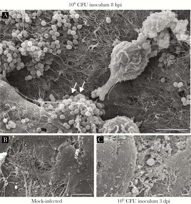
Enterococcus faecalis forms microcolonies in acutely infected wounds. Mice were wounded and infected with 106 CFU E faecalis OG1RF or mock infected with phosphate buffered saline. Wounds were harvested at the indicated postinfection time points for scanning electron microscopy. E faecalis microcolonies encapsulated by fibrous material were visible at 8 hours postinfection (hpi) (white arrows, A), but not in mock-infected wounds (B) or in infected wounds at 3 days postinfection (dpi) (C). Bar represents 5 μm. Images shown are representative of 3 independent experiments.

