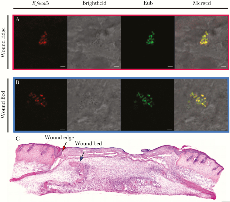
Figure 5.
Enterococcus faecalis is present at the wound edge and in the wound bed. Male C57BL/6 mice were wounded and infected with 106 CFU of E faecalis OG1RF. Wounds were harvested at 3 days postinfection, cryosectioned, and subjected to (A,B) fluorescence in situ hybridization or (C) H&E staining. A, E faecalis-specific probe or probe specific for the domain bacteria (Eub) were used for fluorescence in situ hybridization. The brightfield channel is represented in grey scale. Red and blue arrows (C) correspond to the red and blue boxes (A,B) and represent the wound edge and wound bed, respectively. A,B, Bar represents 2 μm. Images shown are representative of 3 independent experiments.

