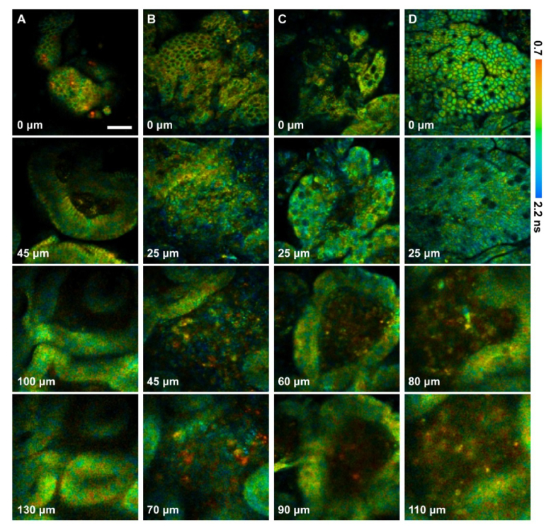
Fig. 8.
Fluorescence lifetime-coded 3D structures of the normal and diseased human gastric antrum mucosa: (A) normal; (B) chronic gastritis with erosion; (C) chronic gastritis with intestinal metaplasia; (D) intestinal-type adenocarcinoma. The depth is labeled in the bottom left corner of each panel. The scale bar is 50 μm.

