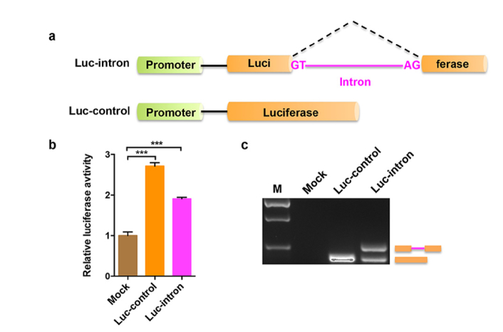
Fig. 1.
Characterization of the splicing reporter. (a) Schematic diagram of the intron-containing (Luc-intron) and intronless (Luc-control) luciferase reporters. Both luciferase genes are under the control of a CMV promoter. GT and AG represent the 5′ and 3′ splice site of intron, respectively. (b) The A549 cells were transfected with the Luc-intron or Luc-control plasmid, together with an internal control plasmid pRL-TK that expresses renilla luciferase. Mock indicates that the cells were added only transfection reagent. Dual luciferase reporter assay was performed 24 h after transfection. Error bars represent the standard deviations for three independent experiments. ***p < 0.001 (c) RT-PCR analysis of total RNA isolated from the cells treated by the same method as described in (b). M indicates DNA marker.

