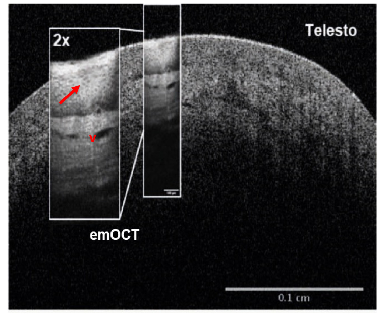
Fig. 5.
Comparison of a 1300 nm OCT image with 10 µm lateral resolution to the emOCT imaging (inset) taken from the same tissue at different positions. Both images of nasal stratified squamous epithelium in the vestibule of the nose were taken in vivo and scaled to the same size. emOCT visualizes otherwise barely visible vessels (v) with high quality and even single cells can be seen (arrow).

