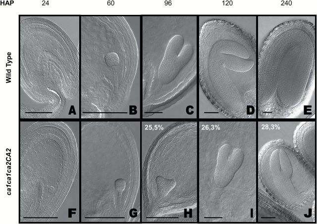
Fig. 2.
Embryogenesis in WT and ca1ca2 mutants. WT (A–E) and double mutant ca1ca1ca2ca2 embryo developmental stages (F–J). The double mutant embryos show a growth delay, perceptible from heart stage (H) of embryogenesis. At the end of embryogenesis, these mutants are at linear cotyledon stage (J) while WT embryos are in mature green stage (E). Images were taken by DIC microscopy on embryos previously cleared 16h in Hoyer’s solution. Percentages in panels H, I and J indicate proportion of embryos in those stages, within a ca1ca1ca2CA2 silique. H, n= 396; I, n= 290; J, n= 435. Bars, 50 μm. HAP, hours after pollination.

