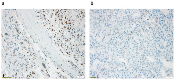
Figure 1.
Different SALL4 staining patterns in HCC. (a) Diffuse nuclear SALL4 staining pattern was seen in 3 cases. These cases were defined as SALL4-positive. (b) Granular nuclear staining pattern was seen in 20 cases. Immunohistochemistry performed on the tumor blocks of these cases confirmed that these cases were SALL4-negative. Scale bar: 1 mm.

