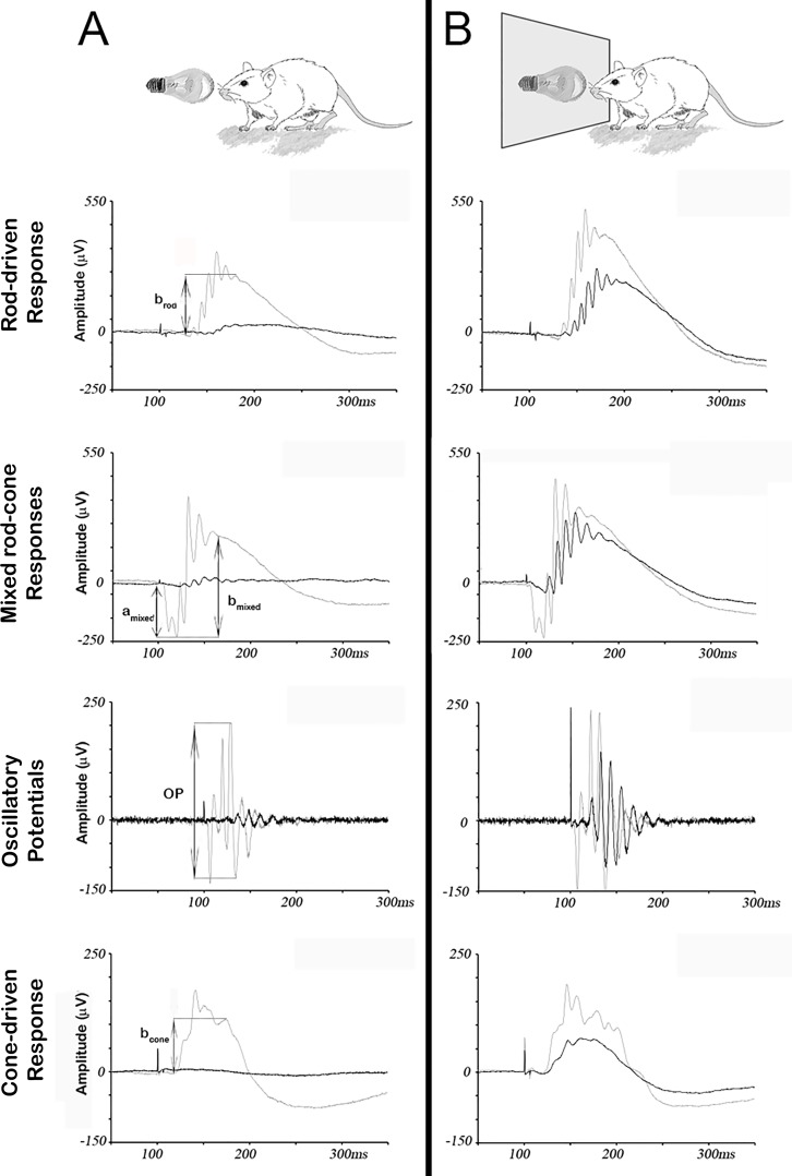
Fig 2. Protective effect of the blue-blocking filter on retinal response to damaging light.
Representative ERG traces from unprotected (A) and protected (B) groups of mice, obtained before (grey traces) and after (black traces) exposure to white light (5,000 lux) for seven days. Arrows indicate the amplitudes measured for each different ERG wave. In the unprotected group (A), amplitudes of scotopic (rod-driven, mixed rod-cone and Oscillatory Potentials) and photopic (cone-driven) parameters are strongly reduced upon light-induced damage, while in the protected group (B), this reduction is attenuated.

