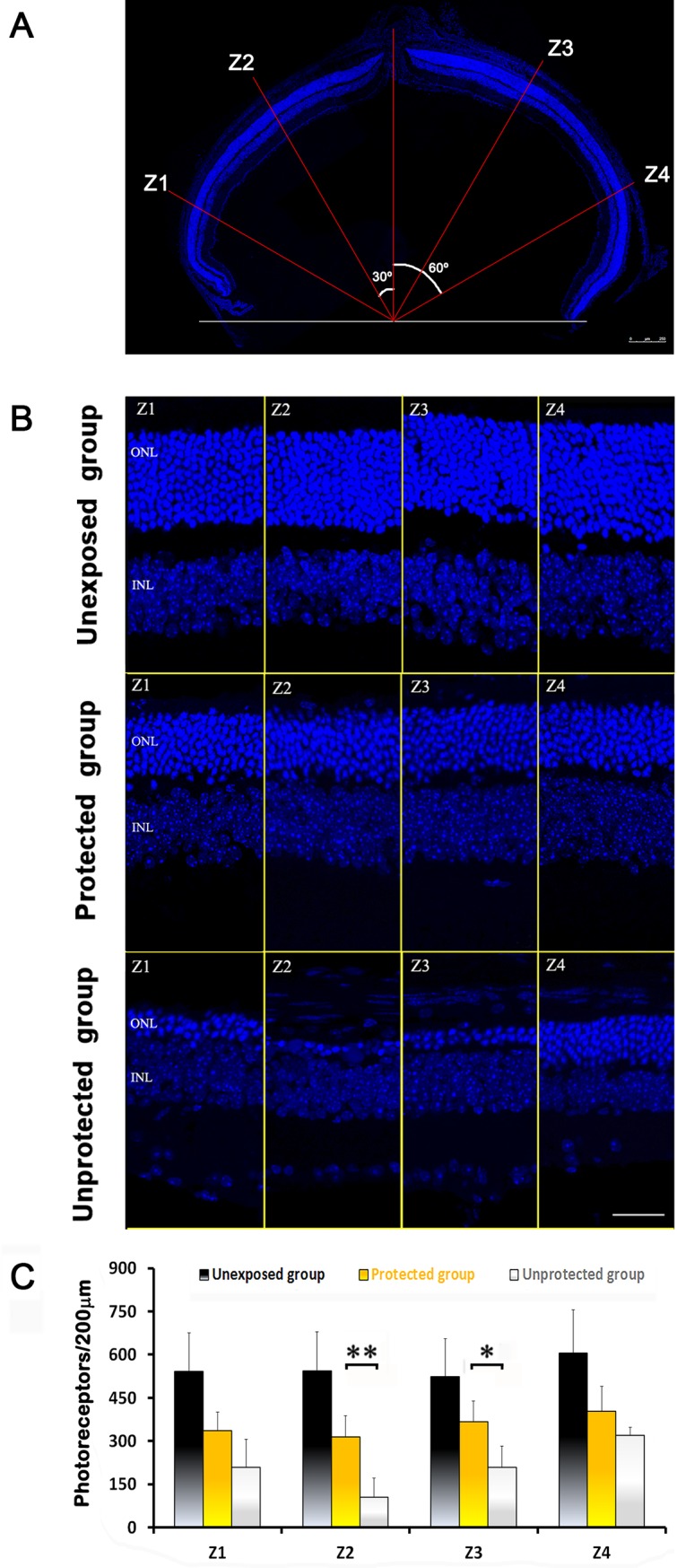
Fig 4. Number of surviving photoreceptors under different experimental lighting conditions.

(A) Photoreceptor survival was analyzed in cryosections at four retinal eccentricities: Z1, 60° dorsal from the optic nerve head (ONH); Z2, 30° dorsal from the ONH; Z3, 30° ventral from the ONH; and Z4, 60° ventral from the ONH. Scale bar 250 μm. (B) Representative images of retinal sections stained with DAPI from the three experimental groups. ONL: outer nuclear layer, INL: inner nuclear layer. Scale bar: 80 μm. (C) Averaged number of photoreceptors (mean ± SD; n = 4) at the different eccentricities. Statistically significant differences were observed between the unexposed group and the other two for all retinal eccentricities (p<0.01, two way ANOVA) and between the unprotected and the protected groups for Z2 and Z3 eccentricities (*p<0.05, **p<0.01, respectively; two way ANOVA).
