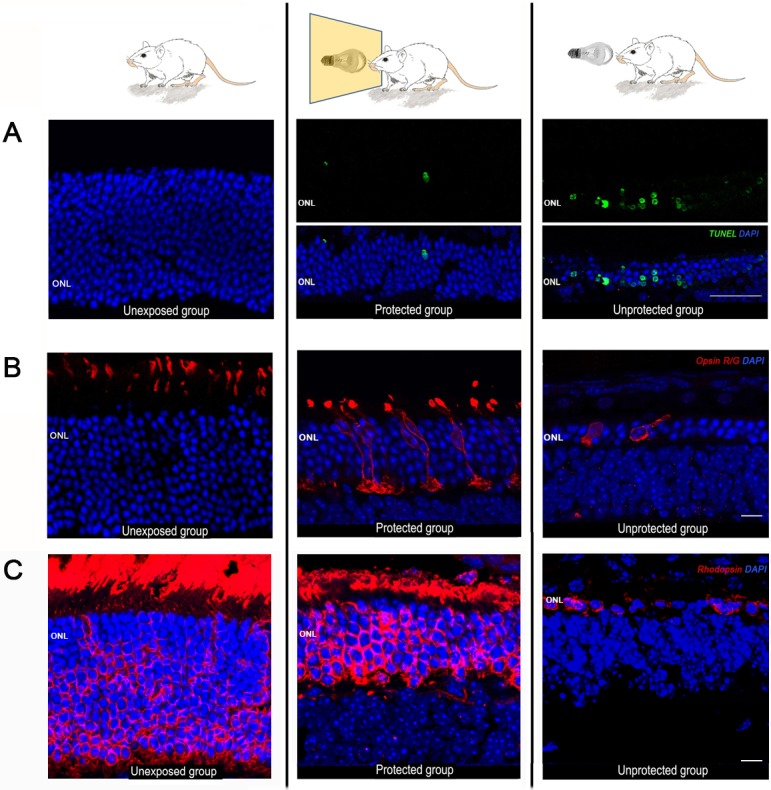
Fig 5. Photoreceptor cell death and protective effect of the blue-blocking filter on cone photoreceptors.

Representative photomicrographs of retinal sections (eccentricity Z2, 30° dorsal from the ONH) obtained from the three experimental groups: unexposed (left panel), protected (middle panel) and unprotected (right panel). (A) Photoreceptor cell death was revealed by the TUNEL technique (green fluorescence). Unprotected animals showed an intense labeling at the ONL, while little or no labeling was observed in protected and unexposed animals, respectively. Nuclei were counterstained with DAPI (blue fluorescence). Scale bar: 50 μm. (B) Immunostaining of cone photoreceptors with red/green opsin antibodies (red fluorescence). Scale bar: 10μm. (C) Immunostaining of rod photoreceptors with rhodopsin antibodies (red fluorescence) Gain settings were maintained at the same level for all experimental groups. Scale bar: 7 μm.
