Figure 1.
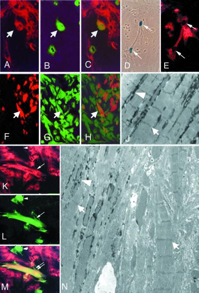
In vitro cardiac differentiation of endothelial cells. (A–C) Double fluorescence of a coculture of neonatal rat cardiomyocytes, stained with anti-MyHC MF20 (red in A) with endothelial progenitors labeled with a GFP lentiviral vector (green in B). Arrows indicate a double-labeled cell (orange in the merged figure, C). (D and E) Coculture of endothelial progenitors, freshly isolated from MLC1/3F-nLacZ day 9 embryos with neonatal rat cardiomyocytes, stained with X-Gal (D) and with anti-cardiac troponin I (E). Arrows indicate a binucleated cell expressing β-galactosidase in the nucleus and cardiac troponin I in the cytoplasm. (F–H) Double fluorescence of a coculture of neonatal rat cardiomyocytes, prelabeled with 6′-carboxyfluorescein (green in G), with endothelial progenitors labeled with an adenoviral vector expressing lacZ and stained with an anti-β-galactosidase antibody (red in F). Arrows indicate one of several endothelial cells where fluorescein has entered through open junctions (orange in the merged figure, H). (K–M) Double fluorescence of a coculture of neonatal rat cardiomyocytes, stained with anti-MyHC MF20 (red in K) with HUVEC labeled with a GFP lentiviral vector (green in L). The double arrow indicates one double-labeled cell (orange in the merged figure, M), whereas the arrowhead indicates a HUVEC that has not trans-differentiated. (N) Electron micrograph showing two close cells, both containing sarcomeres (arrows), one of which is also labeled with Bluo-Gal (arrowhead), whereas the other is not (×3,000). (J) Higher magnification (×5,000) of an area of N of a cell showing Bluo-Gal labeling (arrowhead) among sarcomeres (arrow).

