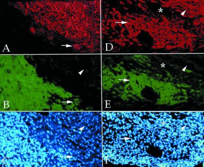
Figure 2.
In vivo cardiac differentiation of endothelial cells. Double fluorescence of a section of normal mouse heart (A–C) and of an infarcted mouse heart (D–F), 2 weeks after injection of endothelial progenitors labeled with a GFP lentiviral vector (green in B and E). The sections have been stained with anti-MyHC MF20 (red in A and D). Nuclear staining (Hoechst) is shown in C and F. In the uninjured heart, very few double-labeled cells could be detected (arrows in A–C). In contrast, a large number of double-labeled cells were present in the infarcted heart (arrows in D–F) in the area of injection; arrowheads indicate normal myocardium, and asterisks indicate an infarcted area devoid of injected cells.

