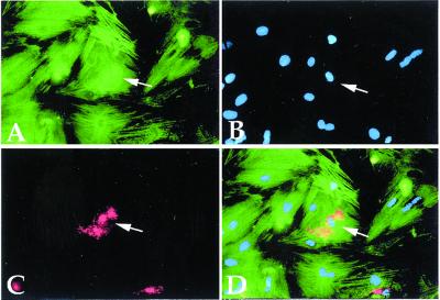
Figure 4.
In vitro cardiac trans-differentiation of endothelial cells. Double fluorescence of a coculture of neonatal rat cardiomyocytes with HUVEC, stained with anti-MyHC polyclonal antibody (green in A), with anti-von Willebrand factor monoclonal antibody (red in C). Nuclear staining (Hoechst) is shown in B. The arrow indicates von Willebrand factor-containing granules in the cytoplasm of a differentiated cardiomyocyte (orange in the merged figure, D).

