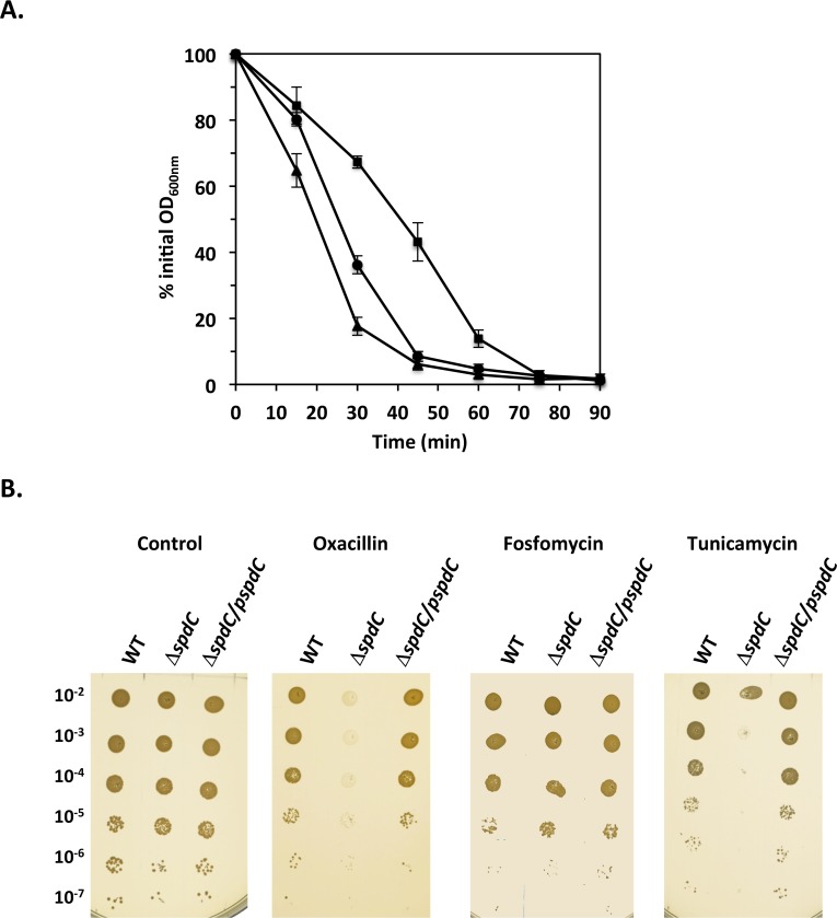
Fig 4. SpdC impacts cell wall homeostasis.
A. The ΔspdC mutant displays increased resistance to lysostaphin-induced lysis. Cells were grown in TSB until mid-exponential phase, harvested and incubated in PBS with lysostaphin (200 ng/ml) with aeration at 37°C. Bacterial lysis was measured by monitoring OD600nm over time. Results are shown as the mean and standard deviation of three independent assays. HG001 parental strain ( ); ΔspdC mutant (■); ΔspdC/pMK4Pprot-spdC complemented strain (▲). B. The absence of SpdC leads to sensitivity to oxacillin and tunicamycin. Dilution series of the HG001, ΔspdC and ΔspdC/ pMK4Pprot-spdC strains on TSA plates with or without antibiotics. Oxacillin: 0.1 μg/ml; fosfomycin: 4 μg/ml; tunicamycin: 1 μg/ml.

