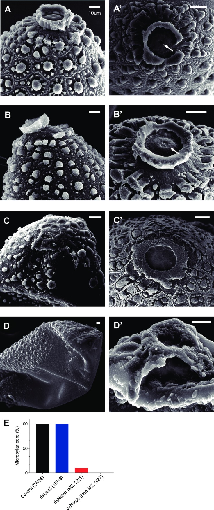
Fig 2. The effect of AaNotch on the ultrastructure of mosquito eggs.

Mosquito eggs from control (A and A’), dsLacZ (B and B’), and dsNotch-treated (C, C’, D and D’) collected at 5 d after egg laying. Arrow: micropylar pore. Scale bar = 10 μm. (E) The percentage of eggs with complete micropylar pore formation. Number in parentheses denotes the number of eggs with complete micropylar pores divided by total number of eggs examined.
