
Figure 1.
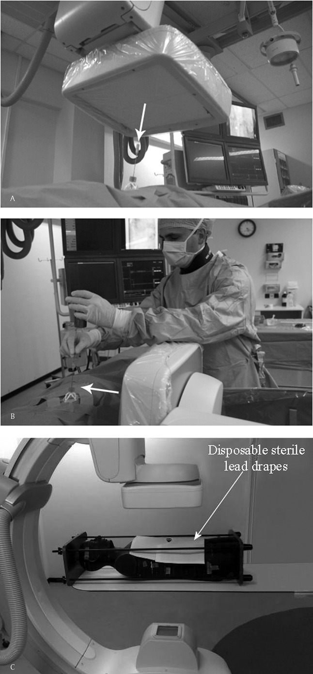
Cone-Beam CT-guidance procedure on a 59-years-old male with a suspected mass in the hilum of the right kidney. Histopathological result revealed a small cell neuroendocrine carcinoma for which this patient underwent a nephrectomy. (A) Entrypoint (EP) geometric configuration of the C-arm, looking ‘down-the-barrel’ (with arrow). (B) shows the common geometric configuration of the C-arm in the Progress view (PV). This clearly shows the close position of the operator to the radiation beam.