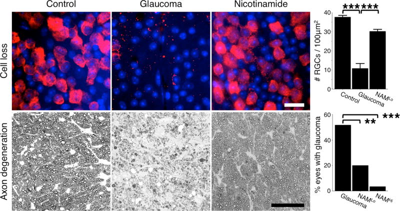
Figure 1. Retinal ganglion cell protection following nicotinamide treatment in the DBA/2J mouse model of inherited glaucoma.
Nicotinamide profoundly protects retinal ganglion cells and prevents optic nerve degeneration in a dose dependent manner. Top row shows flat-mounted retinas stained with an anti-RBPMS antibody that specifically labels retinal ganglion cells (red) and counterstained with DAPI that stains nuclei (blue). There is a significant loss of retinal ganglion cells following periods of elevated IOP (top row, middle panel), which is prevented by nicotinamide treatment (top row, right panel). Bottom row shows cross sections of the optic nerve stained with PPD. Following periods of elevated IOP retinal ganglion cell axons in the optic nerve degenerate and glial scars are formed (bottom row, middle panel). Nicotinamide treatment (NAMLo) robustly protected the axons, and the number of optic nerves with glaucoma was significantly decreased. At a higher dose (NAMHi) 93% of optic nerves did not develop glaucoma (bottom row, chart). Scale bars = 20μm (top row), 50μm (bottom row). ** = P < 0.01, *** = P < 0.001, Student’s t-test (top), Fisher’s exact test (bottom). All images are for mice that were 12 months of age. See references 23, 24 for more details.

