
Fig 5.
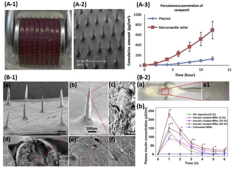
Solid (A) and Porous (B) Microneedles. (A-1) DermaRoller with 540 MNs. (A-2) Side view of SMNs. (A-3) Passive diffusion and MN-mediated diffusion of verapamil with MN rollers across pig skin. Adapted from Kaur et al. [195]. Copyright 2014 Elsevier. (B-1) SEM images of PMNs with different magnifications (a, 500 μm; b, 200 μm), surface region (c,10 μm), and cross-sections (d-f) at different magnifications of 500, 10,000 and 30,000 folds. (B-2) Insulin delivery using PMNs during the treatment process in diabetic rats (a) and plasma insulin levels after administration of insulin-loaded PMN patches (b). Adapted from Yu et al. [206]. Copyright 2014 Elsevier.