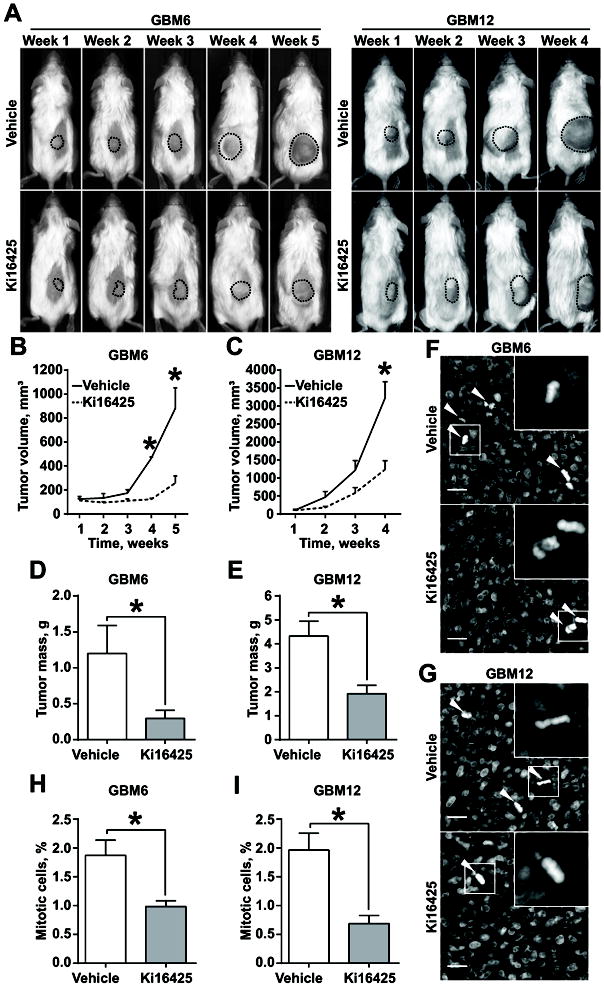
Fig. 6. Inhibition of LPA signaling suppresses proliferation of GBM PDXs in vivo.

(A) Representative images of mice subcutaneously injected with GBM6 (left panel) and GBM12 (right panel) and administered with vehicle or 30mg/kg/day Ki16425. (B, C) Analysis of tumor growth as in (A) for GBM6 (B) and GBM12 (C); 5 mice per group; two-way ANOVA with Sidak’s post hoc test, p<0.05. (D, E) Analysis of GBM6 (D) and GBM12 (E) terminal tumor weight; 5 tumors per group; Student’s t-test, p<0.05. (F, G) Representative images of GBM6 (F) and GBM12 (G), stained with DAPI; arrowheads indicate mitotic cells; scale bar – 20μm. (H, I) Quantification of mitotic figures as in (F, G) for GBM6 (H) and GBM12 (I); at least 1000 cells within 10 random fields per group; Student’s t-test, p<0.05.
