
Figure 1.
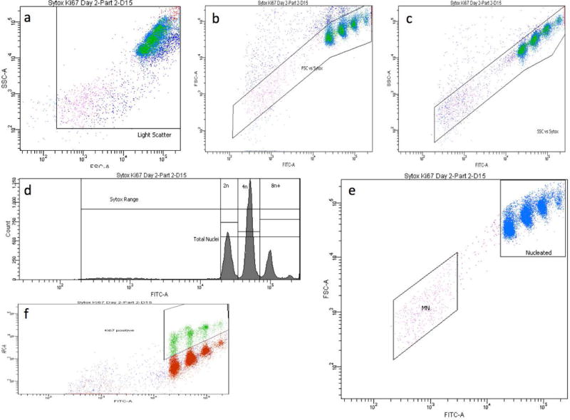
a-f. Bivariate plots and a histogram of liver nuclei and micronuclei from a rat treated with 20 mg DEN/kg/day for 14 days. These panels illustrate the gating strategy used to score the frequency of micronuclei, with simultaneous assessments of hepatocyte proliferation. In order for events to be scored as nuclei or micronuclei, they needed to meet each of the following 4 criteria: within a side scatter vs. forward scatter region, panel a; within a forward scatter vs. SYTOX Green fluorescence region, panel b; within a side scatter vs. SYTOX Green fluorescence region, panel c; and within a SYTOX Green range marker, panel d. Panel d also shows histogram markers used to enumerate the proportion of nuclei with 2n, 4n, and 8n+ ploidy status. Panel e shows the micronucleus-scoring region (MN), which was set to approximately 1/100 to 1/10 the SYTOX fluorescence intensity of G1 nuclei. Panel f shows the region used to enumerate the proportion of nuclei that exhibit anti-Ki-67-associated fluorescence.