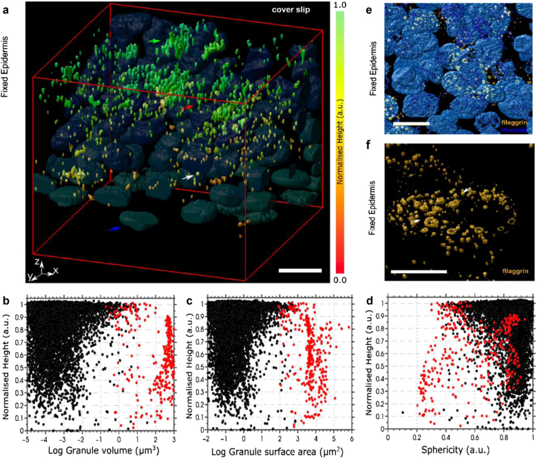
Fig. 1. Filaggrin-containing keratohyalin granules undergo shape alterations and aggregation during formation of stratified epidermis.
a 3D scanning confocal images of fixed epidermal sheets from healthy donors harvested by dispase isolation and stained with anti-filaggrin antibody (G-20 primary and Alexa Fluor 488 secondary antibody). The color scale from red to orange depicts the normalized height relative to the bottom of the epidermis (the most upper epidermal layers at the microscope cover glass in green). Nuclei (blue and representative blue arrow) visualized by Hoechst. The red arrow indicates a representative elongated granule in the mid-layer. The green arrow indicates a representative granule aggregate in the uppermost layer. Scale bar 10 µm; microscope cover glass at the top. The data are representative of eight donors. b–d Geometric analysis of granule distribution within the epidermis based on topological parameters: volume (b), surface area (c), and sphericity (1 marks round granules while values <1 indicate elongated shapes) (d) relative to normalized height (relative to the bottom of the epidermis). Black dots indicate granules smaller than 3 µm in axial length and red dots indicate granules larger than 3 µm in length; n = 9170 granules in 120 cells in N = 8 donors. e, f Magnifications of 3D scanning confocal images of epidermal sheets (as in a) highlighting the morphology of ring-shaped keratohyalin granules (orange; arrows): e scale bar 10 µm; f scale bar 5 µm and nuclei stain removed for clarity. The figure is representative of five donors

