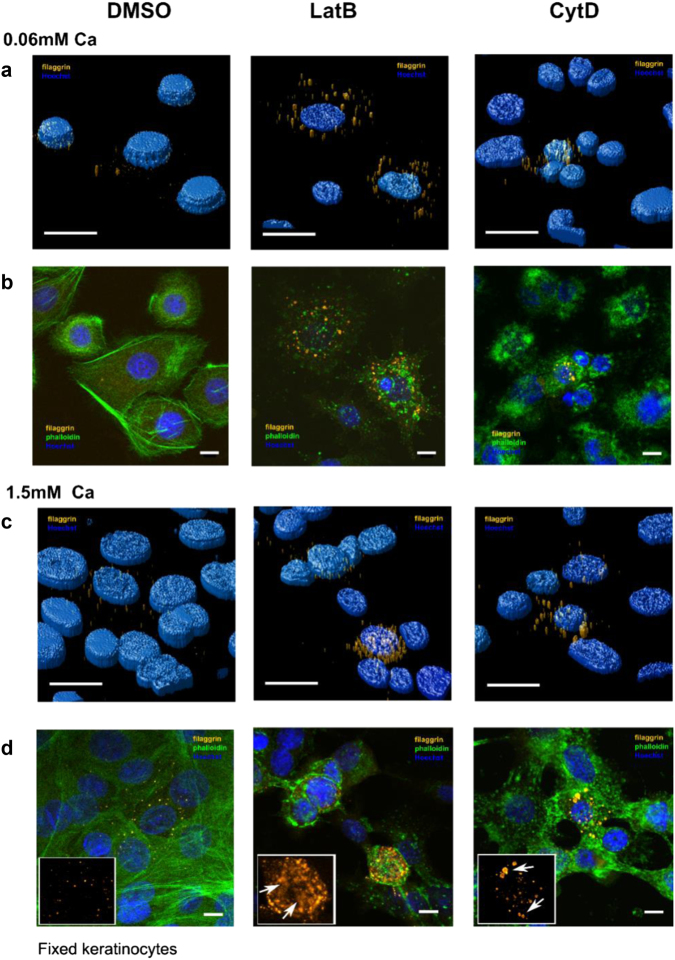
Fig. 5. Actin inhibition increases filaggrin expression but disrupts granule integrity.
Scanning confocal images of fixed NHEKs immunostained for filaggrin (orange, G-20 antibody and Alexa Fluor 488 secondary antibody), F-actin (green, phalloidin-488), and nuclei (blue, Hoechst) without treatment (left panels), with latrunculin B (LatB, middle panels), and with cytochalasin D (CytoD, right panels) treatment after 24 h of treatment with (a, b) [Ca2+] = 0.06 mM and (c, d) [Ca2+] = 1.5 mM. a, c 3D deconvolved images and b, d 2D single X–Y plane images. Insets in d: close-ups and arrows visualize intact granules (right panel) or filaggrin+ clouds (middle panel). Scale bars 10 µm. Data are representative of four separate experiments

