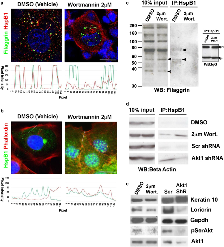
Fig. 6. AKT1-dependent switch between HspB1 interaction with actin and HspB1 interaction with filaggrin and filaggrin processing.
a, b Confocal microscopy of fixed postconfluent rat epidermal keratinocytes treated with vehicle (DMSO) or 2 μm of wortmannin and stained for nucleus (blue, DAPI) and a filaggrin (green, Alexa Fluor 488) and HspB1 (red, Alexa Fluor 594) or b HspB1 (green, Alexa Fluor 488) and actin (red, TRITC–phalloidin). The graphs in each figure show pixel intensity for the red and green channels, respectively, along the line indicated in the micrographs. Scale bar 10 μm. c, d Co-immunoprecipitation of c HspB1 and filaggrin and d HspB1 and β-actin in postconfluent rat epidermal keratinocytes treated with DMSO or wortmannin (Wort); or expressing scrambled or Akt1 shRNA as labeled. 10% Input shows total c filaggrin or d actin (data represent biological replicates: two separate IP experiments). IgH and IgL are IgG heavy and light chains which serve as a loading control for the immunoprecipitation. Arrowheads show the processed filaggrin species altered by wortmannin treatment, and the filaggrin intermediates that are immunoprecipitated by HspB1. e Western blot of keratin-10, loricrin pSer 473 Akt, and AKT1, in wortmannin treated or AKT1 shRNA knockdown keratinocyte (Akt1 ShR, n = 3). GAPDH serves as a loading control

