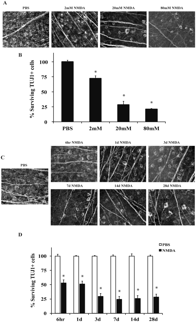
Figure 1.
NMDA induced excitotoxic insult kills RGCs in a dose dependent manner. RGC loss was quantified 7 days after intravitreal injection of NMDA. (A,B) The RGC specific marker, TUJ1, was used to label RGCs in flat-mounted retinas 7 days after a 2 μl intravitreal injection of either PBS (vehicle control) or 2 mM, 20 mM, or 80 mM of NMDA into C57BL/6 J mice (n = 23 for PBS control, n = 6 for 2 mM NMDA, n = 6 for 20 mM NMDA, n = 3 for 80 mM NMDA). Quantification of TUJ1+ RGCs showed NMDA insult caused a dose dependent loss of RGCs. All NMDA concentrations caused a significant decrease in RGC number (*p < 0.001 for all comparisons). Note, that there was no significant difference in RGC number between the 20 mM and 80 mM concentrations (p = 0.819). (C,D) 20 mM NMDA was shown to induce greater than 50% RGC death at 7 days and there appeared to be no benefit to increasing the dose. Thus, 20 mM of NMDA was chosen to investigate the molecular pathways controlling NMDA induced RGC death. To understand the time course of RGC death induced by 20 mM of NMDA, TUJ1+ cells were counted in flat-mounted retinas 6hr, 1d, 3d, 7d, 14d, and 28d after (n ≥ 5 for all time points). NMDA injection resulted in significant loss of RGCs compared to controls at all time points examined (*p < 0.001). Note, approximately 50% of TUJ1 + cells were lost after 6 hours and after 3 days, no more apparent loss of TUJ1+ cells. Scale bar: 50 μm.

