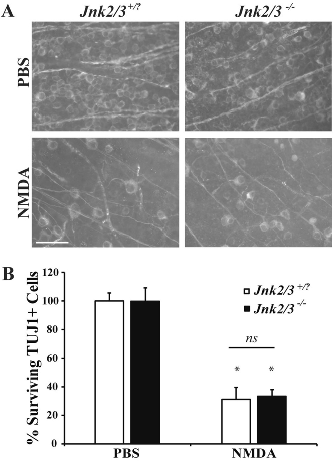
Figure 6.

Jnk2−/−Jnk3−/− nulls do not have attenuated RGC loss after excitotoxic injury. (A) Representative images showing TUJ1 immunolabeled cells in the GCL of Jnk2Jnk3 deficient mice 7 days after intravitreal NMDA or PBS injection, revealing significant RGC loss in both wildtype (Jnk2+/?Jnk3+/?) and Jnk2−/−Jnk3−/− deficient mice after NMDA insult (n = 4 for PBS WT, n = 4 for NMDA WT, n = 3 for PBS Jnk2−/−Jnk3−/−, n = 3 for NMDA Jnk2−/−Jnk3−/−). (B) Quantification of TUJ1 + RGCs confirmed that Jnk2−/−Jnk3−/− deficient mice had a similar loss of RGCs compared to wildtype mice 7d after NMDA injection (*p ≤ 0.001 for comparison between PBS and NMDA; p ≥ 0.9769 comparing WT to Jnk2−/−Jnk3−/− NMDA; ns between genotypes; two-way ANOVA, Tukey’s multiple comparisons test). Scale bar: 50 μm.
