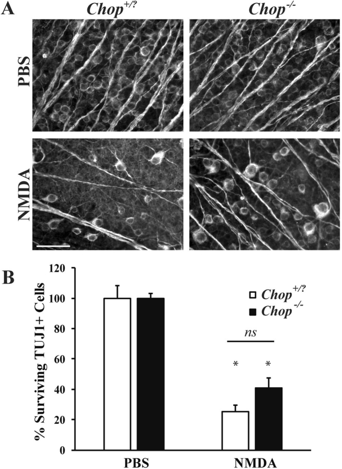
Figure 8.

Ddit3 deficiency provides modest protection of RGCs after NMDA injury. (A) Representative images showing TUJ1 immunolabeled cells in the GCL of Ddit3 deficient (Ddit3−/−), mice 7 days after intravitreal NMDA or PBS injection. Significant RGC loss occurred in wildtype (Ddit3+/?), but Ddit3 deficient mice exhibited modest protection of RGCs after NMDA insult (n = 3 for PBS WT, n = 3 for NMDA WT, n = 8 for PBS Ddit3−/−, n = 10 for NMDA Ddit3−/−). (B) Quantification of TUJ1+ RGCs confirmed that Ddit3 deficient mice had significantly more surviving RGCs compared to wildtype mice 7d after NMDA injection (*p < 0.001 comparing PBS to NMDA; p = 0.8105 comparing WT to Ddit3−/− NMDA; ns between genotypes). Scale bar: 50 μm.
