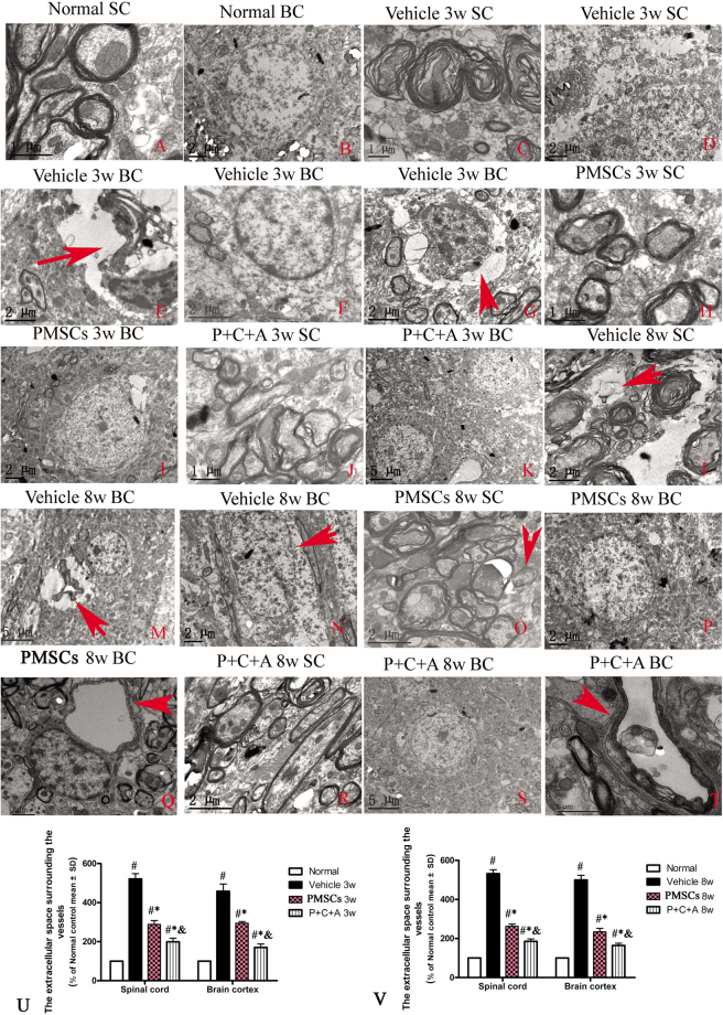
Figure 7.
Electron micrographs demonstrating the prevention of perivascular edema, demyelination/axon loss, and neuronal apoptosis/necrosis in the rat EAE model by PMSC and P + C + A treatment. (A,B) Normal control rats. (A) normal myelinated axons exhibiting dark, ring-shaped myelin sheaths surrounding axons, (B) normal neuronal nuclei with uncondensed chromatin. (C–G) Vehicle-treated EAE rats at 3 weeks pi. (C) myelin sheath displaying splitting, vacuoles, loose and fused changes, and shrunken, atrophied axons, (D,E) tissue edema (D) and severe blood vessel leakage (E, arrow) detected in the extracellular space surrounding the vessels, (F) a neuron showing signs of apoptosis with a shrunken nucleus and condensed, fragmented, and marginated nuclear chromatin, (G) an extravasated inflammatory cell in tissue edema. (H–K) PMSC- (H,I) and P + C + A- (J,K) treated EAE rats at 3 weeks pi. Myelin sheath splitting, axonal loss, and perivascular edema were reduced, and neuron nuclei displayed relatively normal morphology. (L–N) Vehicle-treated EAE rats at 8 weeks pi. Many myelin lamellae were still undergoing vesicular disintegration and demyelination (L), with some fibers being completely lost or only showing an empty circle of remaining myelin (arrow in L). Perivascular edema and leakage as well as extravasated inflammatory cells were still present (M). Some neurons exhibited signs of necrosis such as large vacuoles and degenerated organelles in the perikaryon, ruptured cytoplasmic membranes, and oncolytic chromatins (arrow in N). (O–T) PMSC- (O–Q) and P + C + A- (R–T) treated EAE rats at 8 weeks pi. Newly formed myelin sheaths were detected surrounding intact axons (arrow in O). The morphology of neuron nuclei became relatively normal, especially in the P + C + A-treated group. Perivascular edema and leakage were evidently alleviated. K, M, S, scale bar = 5 µm; B,D,E,F,G,I,L,N,O–R, scale bar = 2 µm; A,C,H,J,T, scale bar = 1 µm. (SC) Transverse sections through the anterior horn of the lumbar spinal. (BC) Coronal sections of the motor cortex. (U,V) The calculations of extracellular space surrounding the vessels at 3 (U) and 8 (V) weeks pi. n = 5, #P < 0.05 vs. normal, *P < 0.05 vs. vehicle, &P < 0.05 vs. PMSCs. (U) In SC, PMSCs vs. vehicle: ES = 0.2, P < 0.0001; P + C + A vs. vehicle: ES = 0.3, P < 0.0001; P + C + A vs. PMSCs: ES = 0.12, P < 0.0001. In BC, PMSCs vs. vehicle: ES = 0.11, P = 0.0003; P + C + A vs. vehicle: ES = 0.17, P < 0.0001; P + C + A vs. PMSCs: ES = 0.32, P < 0.0001. (V) In SC, PMSCs vs. vehicle: ES = 0.51, P < 0.0001; P + C + A vs. vehicle: ES = 0.63, P < 0.0001; P + C + A vs. PMSCs: ES = 0.27, P < 0.0001. In BC, PMSCs vs. vehicle: ES = 0.31, P < 0.0001; P + C + A vs. vehicle: ES = 0.49, P < 0.0001; P + C + A vs. PMSCs: ES = 0.17, P < 0.0001.

