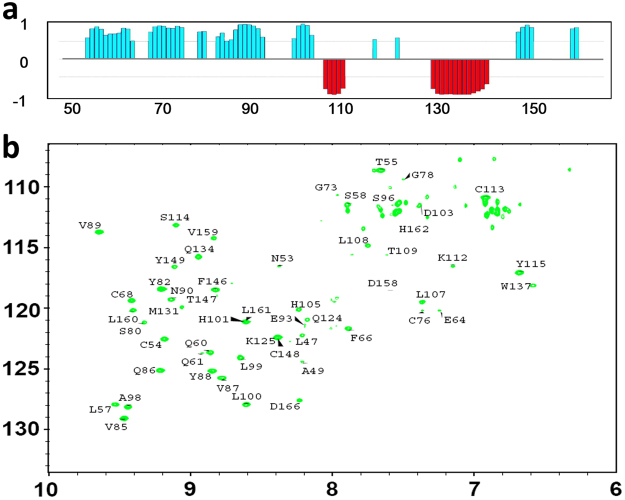
Figure 3.
NMR analysis of secondary structure of hβ4-loop. (a) The chemical shift index (CSI) generated from TALOS program reveals the secondary structure of hβ4-loop. (b) The 1H-15N HSQC spectrum (after the 15N-labeled hβ4-loopsample in H2O NMR buffer was exchanged into D2O NMR buffer) indicates the H-bond formed in α-helices and β-sheets in hβ4-loop, assignments of the cross-peaks belonging to the corresponding residues were displayed.

