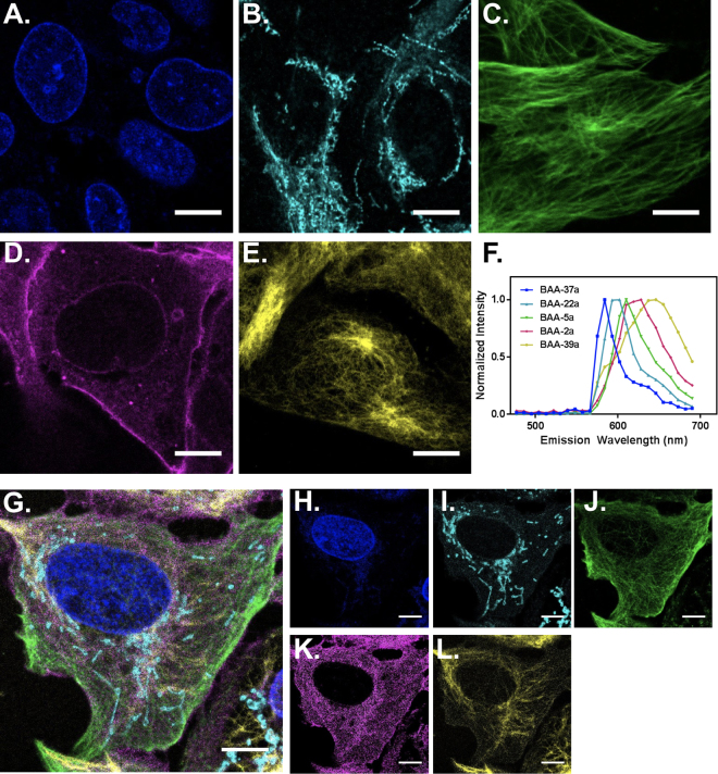
Figure 5.
Immunofluorescence imaging of five subcellular structures. Immunofluorescence imaging using the five selected BAA fluorophores was completed on separate samples to demonstrate the structural and spectral separation of the flurophores and cellular targets. (A) The nuclear membrane was labeled using indirect immunofluorescence with BAA-37a. (B) The outer mitochondrial membrane was labeled using indirect immunofluorescence with BAA-22a. (C) Tubulin was labeled using indirect immunofluorescence with BAA-5a. (D) The extracellular membrane was labeled using BAA-2a conjugated wheat germ agglutinin. (E) Vimentin was labeled using indirect immunofluorescence with BAA-39a. (F) The in situ normalized spectral emission of each BAA fluorophore was collected using the confocal laser scanning microscopy and used for spectral unmixing in (G) simultaneously stained samples created using the same five selected BAA fluorophores conjugated to the validated subcellular tagging reagents. The unmixed, individual spectral channels show the (H) nuclear membrane, (I) mitochondrial membrane, (J) tubulin, (K) extracellular membrane, and (L) vimentin. Scale bar = 10 µm.

