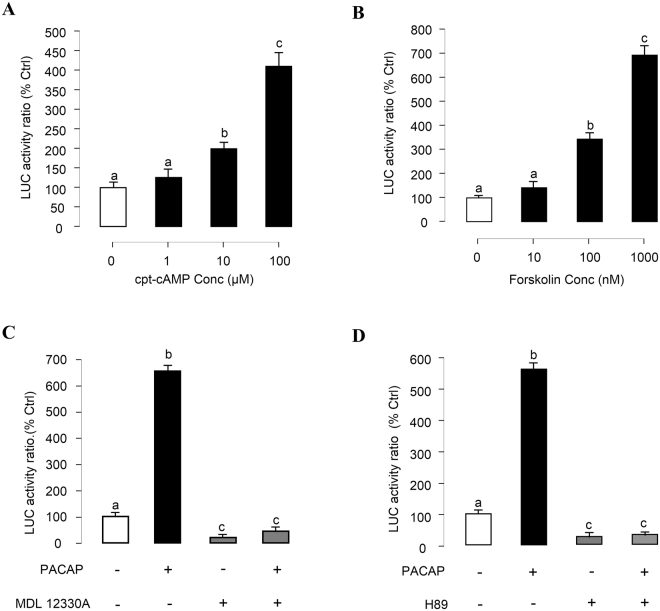
Figure 5.
Functional role of cAMP/PKA pathway in PACAP stimulation of PRL promoter activity in αT3-1 Cells. αT3-1 cells were transiently transfected with pPRL(−1156).LUC for 6 h by using lipofectamine. After 18 h recovery, the cells were incubated with respective drugs. (A) αT3-1 cells over-expressed pPRL(−1156).LUC were treated for 24 hrs with increasing doses of cpt-cAMP (1–100 μM). (B) αT3-1 cells over-expressed pPRL(−1156).LUC were treated for 24 hrs with increasing doses of Forskolin (10–1000 nM). Effects of cAMP/PKA inhibitors on PACAP-induced PRL mRNA expression were then investigated. αT3-1 cells over-expressed pPRL(−1156).LUC were challenged with oPACAP38 (10 nM, 24 hr) in the presence or absence of (C) AC inhibitor MDL12330A (10 μM) or (D) PKA blocker H89 (10 μM). After drug treatment, cell lysate was prepared for dual-luciferase measurement. Data presented were expressed as percentage of control by conversing the ratio of firefly and renilla luciferase in the same sample. Data presented are expressed as mean ± SEM (n = 4) and different letters denote a significant difference at p < 0.05 (ANOVA followed by Fisher’s LSD Test).

