Figure 3.
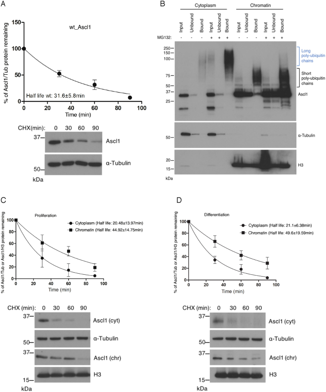
Chromatin-bound Ascl1 is more stable than cytoplasmic Ascl1. (A) P19 cells transfected with (human) Ascl1 and growing in proliferation media were treat with cycloheximide to prevent ongoing protein synthesis. Samples were removed at the times indicated and western blot analysis of whole cell lysates was used to determine Ascl1 half-life. Representative western blots (cropped) are shown and full length western blots are provided in Supplementary Fig. S3A. Error bars = SEM; n = 3 independent experiments. (B) P19 cells transfected with (human) Ascl1 and transferred the day after to differentiation media for 24 hours. Cellular fractionation was performed after 2 hours treatment with or without MG132 and ubiquitin-bound proteins were isolated using the TUBEs method. Western blotting for Ascl1 compared input, resin-unbound and resin-bound fractions. Blue bracket, Ascl1 with long ubiquitin chains; black bracket, Ascl1 with short ubiquitin chains. Also shown are α-Tubulin (control for cytoplasmic fractionation) and histone H3 (control for chromatin fractionation). (C) P19 cells were transfected with (human) Ascl1 and treated the day after with cycloheximide for increasing times before sample withdrawl, as shown. Cytoplasm and chromatin fractions were processed for Ascl1 western blot analysis to determine Ascl1 half-life. Representative western blots (cropped) are shown below and full length western blots are provided in Supplementary Fig. S3B. Error bars = SEM; n = 3 independent experiments. (D) As (C) but with P19 cells transferred to differentiation media for 24 hours prior to cycloheximide addition. Full length western blots are provided in Supplementary Fig. S3C.

