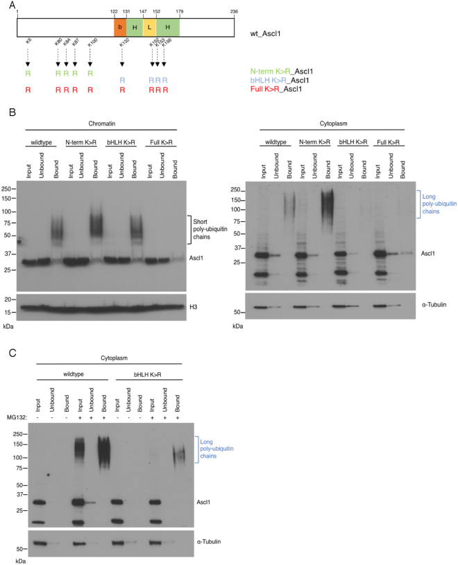
Figure 5.
Sites available for ubiquitylation differ in cytoplasmic and chromatin bound Ascl1. (A) Schematic representation of human Ascl1 indicating mutation of lysines into arginines at the N-terminus and within the bHLH domain. (B) P19 cells transfected with wild-type Ascl1, N-term K > R_Ascl1, bHLH K > R_Ascl1 or Full K > R_Ascl1 were transferred the day after to differentiation media for 24 hours. Cellular fractionation was performed, and ubiquitylated proteins were isolated using the TUBEs method. Western blotting for Ascl1 compared input, resin-unbound and resin-bound fractions from cytoplasmic and chromatin compartments as labelled. Black bracket, Ascl1 with short ubiquitin chains in the chromatin fraction; blue bracket, Ascl1 with long ubiquitin chains in the cytoplasmic fraction. Also shown are α-Tubulin (control for cytoplasmic fractionation) and histone H3 (control for chromatin fractionation). (C) As (B) above, except cells were incubated with or without MG132 before harvesting to inhibit ubiquitin-mediated proteolysis. Western blots for loading control were cropped to show specific band of correct molecular weight.

