Abstract
Plasmodiophora brassicae is a soil-borne pathogen that belongs to Rhizaria, an almost unexplored eukaryotic organism group. This pathogen requires a living host for growth and multiplication, which makes molecular analysis further complicated. To broaden our understanding of a plasmodiophorid such as P. brassicae, we here chose to study immunophilins, a group of proteins known to have various cellular functions, including involvement in plant defense and pathogen virulence. Searches in the P. brassicae genome resulted in 20 putative immunophilins comprising of 11 cyclophilins (CYPs), 7 FK506-binding proteins (FKBPs) and 2 parvulin-like proteins. RNAseq data showed that immunophilins were differentially regulated in enriched life stages such as germinating spores, maturing spores, and plasmodia, and infected Brassica hosts (B. rapa, B. napus and B. oleracea). PbCYP3 was highly induced in all studied life stages and during infection of all three Brassica hosts, and hence was selected for further analysis. PbCYP3 was heterologously expressed in Magnaporthe oryzae gene-inactivated ΔCyp1 strain. The new strain ΔCyp1+ overexpressing PbCYP3 showed increased virulence on rice compared to the ΔCyp1 strain. These results suggest that the predicted immunophilins and particularly PbCYP3 are activated during plant infection. M. oryzae is a well-studied fungal pathogen and could be a valuable tool for future functional studies of P. brassicae genes, particularly elucidating their role during various infection phases.
Electronic supplementary material
The online version of this article (10.1007/s00438-017-1395-0) contains supplementary material, which is available to authorized users.
Keywords: Cyclophilin, Immunophilin, Plasmodiophora brassicae, Rhizaria
Introduction
The plant immune system recognizes microbial pathogens in various ways to prevent infection. Pathogens try on the other hand to evade recognition and defense responses in plants. Knowledge on these events has rapidly increased over the last years (Boutrot and Zipfel 2017). However, pathogens with obligate biotrophic lifestyle are lagging behind in this context, since extraction of high-quality nucleic acids and any kind of gene editing are difficult and in some cases almost impossible. Plasmodiophora brassicae is a good example of this type of category. This organism is an obligate biotrophic plasmodiophorid that belongs to the Phytomyxea class within the eukaryote supergroup Rhizaria, taxonomically distinct from other plant pathogens, such as fungi or oomycetes (Neuhauser et al. 2011; Sierra et al. 2016). Rhizaria is one of the least studied groups of eukaryotes (Sibbald and Archibald 2017). Besides P. brassicae, few other genomes are available in Rhizaria; all for diverse species such as the chlorarachniophyte alga Bigelowiella natans, the foraminifera Reticulomyxa filosa, the transcriptome of the potato powdery scab pathogen Spongospora subterranea and few transcriptome datasets on marine species (Curtis et al. 2012; Glöckner et al. 2014; Keeling et al. 2014; Schwelm et al. 2015; Krabberød et al. 2017). The P. brassicae genome is relatively small (25.5 Mb) compared to the free-living B. natans (~ 100 Mb) and R. filosa (~ 320 Mb). Plasmodiophorids infect a wide range of host organisms (Neuhauser et al. 2014). The Brassicaceae plant family is the preference of P. brassicae, the clubroot disease agent. This disease is increasing in importance, causing a 10–15% yield reduction on a global scale (Dixon 2009). Due to the hidden lifestyle in the soil and its requirement of a living host plant root for growth and multiplication, many aspects on this plant pathogen remain to be elucidated. This also calls for new tools for functional gene assessments.
Immunophilins are ubiquitous proteins with properties that allow them to regulate protein structure, activity and stability (Wang and Heitman 2005; Hanes 2015). They operate either by peptidyl-propyl isomerization of selected targets, as chaperons or by binding of small ligands. The immunophilins comprise of three structurally unrelated subfamilies: the cyclophilins (CYPs), the FK506-binding proteins (FKBPs), and the parvulin-like proteins (Hanes 2015). Besides modulating the formation of cis–trans isomers of proline (Galat 2003), immunophilins have two important amino acid properties: (a) prolyl-isomerase activity, which catalyzes the rotation of the X-Pro peptide bonds from the cis to trans configuration, a rate-limiting step in protein folding (Wang and Heitman 2005), and (b) affinity to bind to immunosuppressive drugs. The stramenopile human parasite Blastocystis sp. lives under anaerobic conditions partly like P. brassicae (Gravot et al. 2016) and secretes a range of immunophilins with potential effector functions, which could lead to cell death (apoptosis) in the host tissue (Denoeud et al. 2011). Another animal parasite, the protozoan Toxoplasma gondii uses a cyclophilin (TgCYP18) to manipulate host cell responses (Ibrahim et al. 2009). Among plant pathogens, some cyclophilins are able to inhibit RNA replication of plant viruses (Lin et al. 2012; Kovalev and Nagy 2013), while some effector proteins interact with plant cyclophilins, and thereby induce plant defense responses (Domingues et al. 2010, 2012; Kong et al. 2015). In the rice blast fungus Magnaporthe oryzae, the cyclophilin A homolog MgCYP1 acts as a virulence determinant. When this gene was inactivated it led to reduced virulence, and the Cyp1 mutant strain was malfunctioned regarding formation of penetration peg and appressorium turgor generation (Viaud et al. 2002). MgCYP1 is also the cellular target for the drug cyclosporin A in M. oryzae, functioning as an inhibitor of appressorium development and hyphal growth in a CYP1-dependent manner, suggesting a role for the calcineurin regulation of appressorium development (Viaud et al. 2002).
This work aimed to monitor the presence of immunophilin encoding genes in the genome of P. brassicae, and to analyze potential candidates for function, here using the molecular amenable plant pathogen M. oryzae as heterologous test system. All with the overall aim of enhancing our understanding of this root gall (club)-inciting plant pathogen.
Materials and methods
Prediction of immunophilins, domain analysis and subcellular localization
To identify putative members of immunophilins in P. brassicae, the Hidden Markov Model profiles, unique to cyclophilin (PF00160), FKBP (PF00254) and parvulins (PF00639) from the Pfam database version 28.0 (Finn et al. 2016), were retrieved and searched against the annotated genome of P. brassicae (Schwelm et al. 2015). Protein candidates were identified as described previously (Singh et al. 2014; Tripathi et al. 2015). Significant hits (e value: 1.0E–0) with positive scores were selected for further classification. In analogy with earlier denominations, the proteins identified were named with the prefix Pb (P. brassicae), followed by CYP (cyclophilin), FKB (FK506-binding proteins), and PAR (parvulin-like proteins), according to the catalytic domain present, followed by numbers in increasing order based on the highest Hidden Markov Model scores. Protein domain structures were confirmed with the SMART software (Letunic et al. 2015).
Subcellular localization of the putative immunophilin proteins was predicted using PSORT (Nakai and Horton 1999). Signal peptide cleavage sites and mitochondrial-targeted peptides were predicted using SignalP 4.1 (Petersen et al. 2011) and the TargetP 1.1 server, respectively (Emanuelsson et al. 2000). Nuclear localization signals (NLS) were predicted using NLS mapper (Kosugi et al. 2009).
Phylogenetic analysis
Phylogenetic analysis was conducted on PbCYP3 homologs, derived from different phytopathogens and the host Arabidopsis thaliana, based on catalytic domain sequences and was carried out using the maximum likelihood method implemented in the MEGA v.7 software (Kumar et al. 2016), using the JTT substitution model (Jones et al. 1992). Bootstrap analysis was performed on 1000 replicates. The CLUSTAL W algorithm was used for alignments (Thompson et al. 1994) (Supplementary dataset 1).
Transcriptome analysis
The transcriptome of P. brassicae genes, coding for putative immunophilins, was analyzed exploiting data from various life-stage-specific forms, such as germinating spores, maturing spores and plasmodia of P. brassicae and in clubroot-infected Brassica hosts (B. rapa, B. napus and B. oleracea), as described by Schwelm et al. (2015). Fragments per kilobase of transcript per million mapped reads (FPKM) were calculated (Trapnell et al. 2010). Furthermore, the immunophilin gene expression levels were visualized in heat maps as log10-transformed FPKM values and were normalized by calculating the Z-score for each gene across all transcriptome libraries. Heat maps were drawn using the gplot package of R software (R Core Team 2014), as described previously by Singh et al. (2014).
Construction of overexpression vector and Magnaporthe oryzae transformation
The P. brassicae single spore isolates e3, was used for spore isolation and purification (Fähling et al. 2004). DNA was extracted from purified P. brassicae resting spores using a NucleoSpin® Plant II Mini kit (Macherey–Nagel) following manufacturer’s instructions. PbCYP3 was PCR amplified from genomic DNA using high fidelity Phusion Taq polymerase (Thermo Scientific) and BamHIPbCyp3 and KpnIPbCyp3 primers (Table 1). The pCB1532 vector, conferring resistance to chlorimuron ethyl (Yang and Naqvi 2014), was used as a destination vector to construct the pCB1532-PbCYP3+ overexpression vector. The orientation and integrity of the insertion were confirmed by DNA sequencing (Macrogen Inc.).
Table 1.
List of primers used in the current study
| Primer name | 5′–3′ |
|---|---|
| BamHIPbCyp3 | ATATGGATCCATGGCGAACCCGAAGGTCT |
| KpnIPbCyp3 | ATATCCATGGTCAGCACTCGCCGCACTTC |
| OsElfF | TTGTGCTGGATGAAGCTGATG |
| OsElfR | GGAAGGAGCTGGAAGATATCATAGA |
| MgActF | ATGTGCAAGGCCGGTTTCGC |
| MgActR | TACGAGTCCTTCTGGCCCAT |
| PbCYP3C_RT-F | ATTTCACGAACCACAACGGCACTG |
| PbCYP3C_RT-R | TGGACACGGTGCACACGAAGAAC |
Construction of M. oryzae ΔCyp1+ overexpression strain (containing the P. brassicae PbCYP3 gene) was carried out using a protoplast-mediated protocol. Briefly, M. oryzae mycelia from the ΔCyp1 strain (Viaud et al. 2002), with the Guy11 genomic background, were incubated in OM buffer (1.2 M MgSO4, 10 mM Na-PO4 pH 5.8) containing lytic enzymes (Novozymes). Protoplasts were mixed with the pCB1532-PbCYP3+ overexpression vector in STC buffer (1.2 M sorbitol, 10 mM Tris–HCl pH 7.5, 10 mM CaCl2) and incubated at room temperature for 25 min. PTC buffer (60% PEG 4000, 10 mM Tris–HCl pH 7.5, 10 mM CaCl2) was then added followed by incubation at room temperature for 20 min. Finally, protoplasts were added to molten BDC medium (yeast nitrogen base without amino acids and ammonium sulfate, 1.7 g/L ammonium nitrate, 2 g/L, asparagine, 1 g/L, glucose, 10 g/L, pH 6, 1.5% agar). The plates were incubated for at least 16 h at 24 °C and overlaid with approximately 15 ml BDC medium without sucrose, supplemented with 1.5% agar and 150 μg mL−1 chlorimuron ethyl (Sigma Aldrich). Fungal colonies resistant to chlorimuron ethyl were verified by PCR that contain the PbCYP3 gene, and expression levels of this gene confirmed using RT-qPCR as described below.
Quantitative real-time PCR
Total RNA was extracted from 1-week-old cultures of wild-type (Guy 11), ΔCyp1 and ΔCyp1+ M. oryzae strains using the RNeasy Plant Mini Kit (Qiagen) according to manufacturer’s instructions and concentrations were determined using NanoDrop (Thermo Scientific). For cDNA synthesis, 1 µg total RNA was reversed transcribed in a total volume of 20 µl using the iScript cDNA Synthesis Kit (Bio-Rad). Transcript levels were quantified by quantitative reverse transcriptase PCR (RT-qPCR) using the iQ5 qPCR System (Bio-Rad) as described previously (Tzelepis et al. 2012). Relative expression values of PbCYP3 gene were calculated using the 2−ΔΔCT method (Livak and Schmittgen 2001). The M. oryzae actin gene (Che Omar et al. 2016) was used to normalize the data using the MgActF/R primers listed in Table 1.
Phenotypic analysis and quantification of Magnaporthe oryzae biomass in infected plants
Magnaporthe oryzae Guy11 (WT), ΔCyp1+ and ΔCyp1 strains were grown in triplicates on oatmeal agar plates and kept for 1 week at 25 °C in darkness. Rice plants of the cultivar CO-39 (Oryza sativa) were grown under controlled conditions at 28 °C in cycles of 14 h light and 10 h dark. 4-week-old plants were inoculated with 2 mm mycelia plugs, derived from 2-week old M. oryzae cultures of wild-type, ΔCyp1+ and ΔCyp1, while mock inoculation was conducted with only agar plugs as previously described (Dong et al. 2015). Fungal colonization on leaves were monitored after 1 week and DNA was extracted using a CTAB method (Möller et al. 1992) and quantified using the M. oryzae actin (act) gene, normalized with the elongation factor gene (elf-1) from Oryza sativa (Ma et al. 2011) and qPCR techniques as described above. At least five biological replicates were used. For the statistical analyses, the ANOVA (one way) was conducted using a general linear model implemented in SPSS 20 (IBM). Pairwise comparisons were performed using the Tukey’s method at the 95% significance level (Tukey 1949).
Results
Sequence and structure characteristics of Plasmodiophora brassicae immunophilins
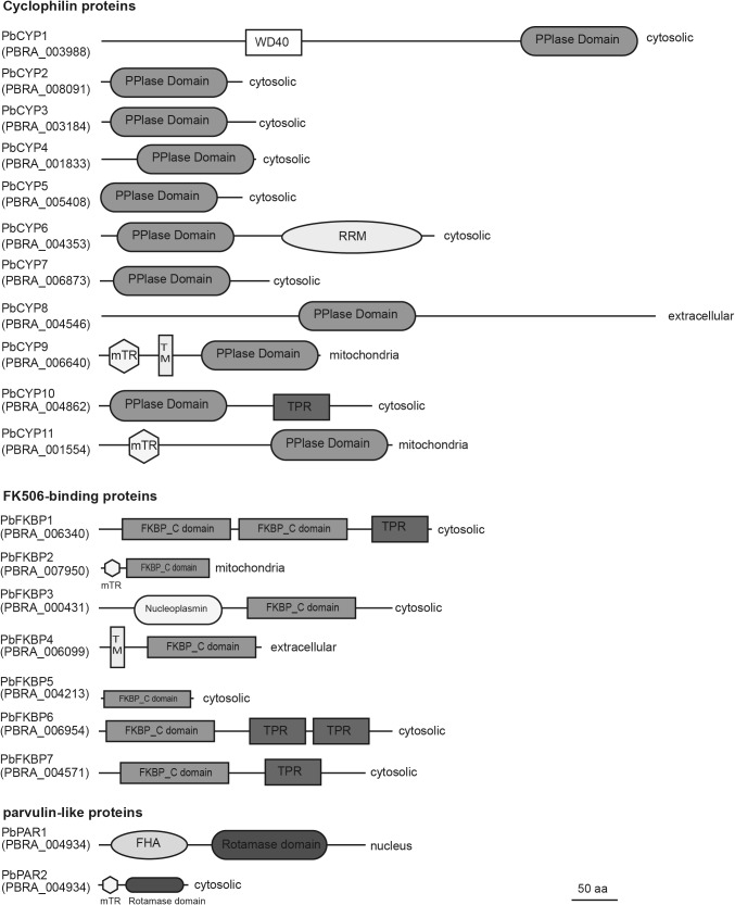
Our analysis revealed that the P. brassicae genome contained 20 genes encoding putative immunophilins (Fig. 1). Eleven belong to the cyclophilins, seven to FKBP and two to the parvulin subfamilies. Seven proteins carried a single catalytic domain, while a transmembrane domain was predicted for two. Only the PbCYP1 protein harbored a WD-40 domain, while a nucleoplasmin domain was detected in the PbFKBP3 sequence. Four proteins harbored tetracopeptide repeats, while PbPAR1 and PbCYP6 each contained a forkhead-associated or a RNA recognition motif. Based on localization signals, following distribution was predicted; PbCYP8 and PbFKBP4 were extracellular, PbPAR1 was localized to the nucleus, while PbCYP9, PbCYP11 and PbFKBP2 were localized to the mitochondria. The remaining 14 proteins predicted to be localized in the cytosol (Fig. 1).
Fig. 1.
Domain architecture of predicted immunophilins in Plasmodiophora brassicae genome. Amino acid sequences were analyzed for the presence of conserved domains using the SMART software. WD40 tryptophan-aspartic acid repeats, PPIase peptidyl-prolyl isomerase, RRM RNA recognition motif, FKBP FK506-binding protein, TPR tetracopeptide repeat, FHA forkhead-associated domain, rotamase domain parvulin-like rotamase domain, mTP mitochondrial target peptide, TM transmembrane domain. Predicted cell localization is indicated. The bar marker indicates a length of 50 amino acids and refers to total protein length
Expression patterns of immunophilin genes in various P. brassicae stages and clubroot-infected plants
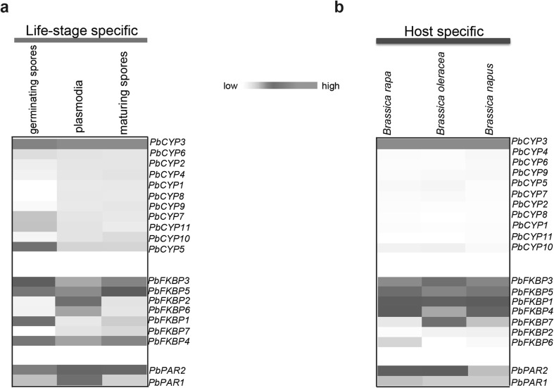
Re-analyzed RNAseq data from Schwelm et al. (2015) generated differential expression patterns for genes, encoding for putative immunophilins, in various enriched life stages, such as germinating and maturing spores, plasmodia and clubroot-infected Brassica hosts (B. rapa, B. napus and B. oleracea). Notably, PbCYP3 was highly induced in all studied life stages (Fig. 2a), while PbCYP5, PbCYP7 and PbCYP11 showed elevated transcript levels in germinating spores. The other PbCYP genes had low transcript levels. Only the PbCYP3 gene displayed high induction during infection of the three Brassica hosts, while others showed low expression levels (Fig. 2b). In the FKBP subfamily, PbFKBP3 and PbFKBP5 were highly activated during different life stages. PbFKBP2 and PbFKBP6 were upregulated in plasmodia, and PbFKBP1 during the germinating spore stage (Fig. 2a). Notably, PbFKBP3 and PbFKBP5 were highly induced in all host species, whereas PbFKBP7 was highly induced only in B. oleracea (Fig. 2b). In the case of the PAR subfamily, PbPAR2 was constitutively expressed during all stages compared to PbPAR1 (Fig. 2a). High transcript level of PbPAR2 was also observed in B. rapa and B. oleracea compared to B. napus, while PbPAR1 was expressed at low levels in all Brassica hosts.
Fig. 2.
Expression patterns of the predicted immunophilins (IMMs) from P. brassicae. The heat maps show the RNAseq-based expression profile of IMMs during: a different Plasmodiophora brassicae life stages; germinating spores, plasmodia and maturing spores, and b different clubroot-infected Brassica hosts; B. rapa, B. napus and B. oleracea. The heat maps were drawn using the gplot package of R statistical software
Sequence, phenotypic and pathogenicity analyses of PbCYP3
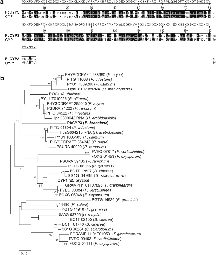
In our sequence comparison, the P. brassicae PbCYP3 cyclophilin showed 60% similarity to the M. oryzae CYP1 (Fig. 3a). In phylogenetic analysis of PbCYP3 homologs present in plant pathogenic fungi and oomycetes, PbCYP3 grouped together with homologs from oomycetes (Fig. 3b), while M. oryzae CYP1 clustered in a separate group together with cyclophilins derived from filamentous ascomycetes.
Fig. 3.
Comparison of Plasmodiophora brassicae PbCYP3 and Magnaporthe oryzae CYP1. a Amino acid alignment between PbCYP3 and CYP1. Sequence similarities are depicted with black shade. Alignments were conducted using the CLUSTALW algorithm implemented in MegAlign software (DNASTAR, Madison). b Phylogeny of homologs to the PbCYP3 cyclophilin. Analysis was conducted using maximum likelihood with the JTT substitution model, based on Clustal W alignment using all amino acid sites. The bar marker indicates the numbers of amino acid substitutions. Protein identifiers include protein name (if available) or protein ID accession numbers from TAIR and FungalDB databases. Plasmodiophora brassicae and Magnaporthe oryzae cyclophilins are indicated in bold. Bootstrapping was with 1000 replicates and values ≥ 50 are shown
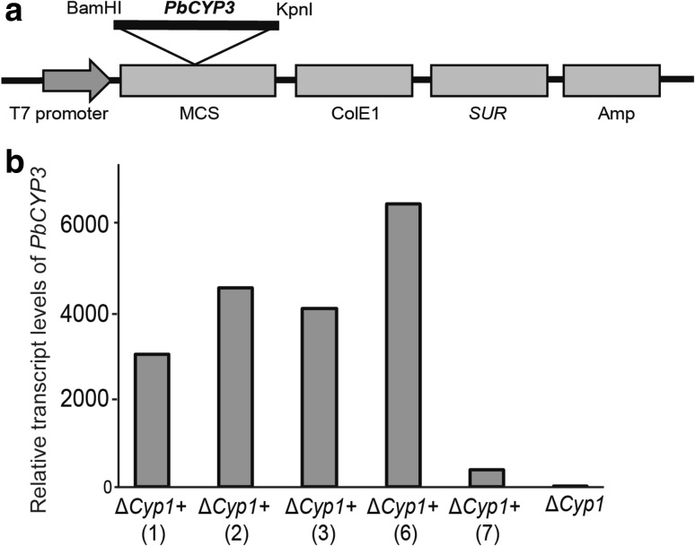
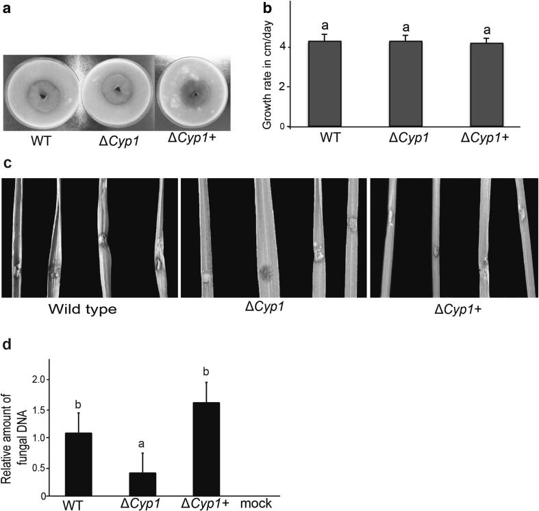
Heterologous expression of the PbCYP3 in the M. oryzae deletion strain ΔCyp1 was produced using the pCB1532:PbCyp3+ vector (Fig. 4a). Five selected chlorimuron ethyl-resistant M. oryzae transformants showed elevated transcript levels of the PbCYP3 gene (Fig. 4b). The ΔCyp1+ transformant (no. 6), which showed the highest transcript levels, was used for further analysis. No difference in colony morphology and mycelial growth rate was observed between M. oryzae Guy11 (wild-type), ΔCyp1, and the ΔCyp1+ strains (Fig. 5a, b). To evaluate the role of PbCYP3 in virulence, rice plants were infected with these three strains independently. Our results showed that the wild-type strain caused more severe symptoms on rice plants compared to symptoms caused by the ΔCyp1 and ΔCyp1+ strains (Fig. 5c). However, no significant differences between wild-type and the ΔCyp1+ strain were observed in fungal biomass 7 days post infection, whereas the ΔCyp1 strain had significantly lower DNA levels, signifying reduced plant colonization ability (Fig. 5d).
Fig. 4.
Construction and validation of Magnaporthe oryzae ΔCyp1+ strain. a Map of pCB1532:PbCyp3+ vector used for M. oryzae transformation. The plasmid confers resistance to chlorimuron ethyl (SUR) in M. oryzae and to ampicillin (Amp) in bacteria. b Expression profiles of the PbCYP3 gene in five (1–7) positive ΔCyp1+ transformants. Relative transcription levels in relation to actin gene (act) expression are calculated from Ct values and according to DDCt method
Fig. 5.
Functional analysis of Magnaporthe oryzae strains. a Colony morphology of M. oryzae Guy11 wild-type (WT), ΔCyp1 and ΔCyp1+ strains grown on oatmeal agar. Cultures were maintained for 2 weeks at 25 °C in darkness. b Growth rate of M. oryzae WT, ΔCyp1 and ΔCyp1+ strains grown on oatmeal agar. c Symptoms of M. oryzae WT, ΔCyp1 and ΔCyp1+ strains on rice plants 7dpi. d Biomass quantification of M. oryzae WT, ΔCyp1 and ΔCyp1+ strains upon infection of rice plants cv. CO-39. DNA was extracted from plants 7 days post infection. For quantitative PCR (qPCR), the M. oryzae actin (act) gene was used and data were normalized with the elongation factor gene (elf-1) from Oryza sativa. Letters (a, b) indicate statistically significant differences (p value < 0.05) according to Tukey’s HSD test. Error bars represent SD based on at least five biological replicates
Discussion
The immunophilin repertoire in P. brassicae comprises 20 putative members, distributed on three subfamilies. This gene family size is also common in other eukaryotes such as nematodes, fungi, and oomycetes (Page et al. 1996; Pemberton 2006; Gan et al. 2009; Krucken et al. 2009; Singh et al. 2017). Commonly, plants harbor 50–60 immunophilin proteins (Vasudevan et al. 2015). Arabidopsis thaliana, a host of P. brassicae has 52 immunophilins (He et al. 2004). Much of immunophilin gene function in plants is associated to development but also to different abiotic and stress responses. The P. brassicae nuclear genome lacks a number of gene coding for essential metabolites (Schwelm et al. 2015), which is expected to be a feature of the poorly understood biotrophic lifestyle where the pathogen has evolved a strict dependency with its host (Kemen et al. 2011). Whether there is any overlapping function between the CYP genes in P. brassicae and those in the host is, as most gene functions in this specific plant–pathogen interaction, not known. The PbCYP3 homolog in A. thaliana is the cyclophilin ROC1 (AT4G38740), a gene known to function in the immune response pathways to Pseudomonas syringae (Aumüller et al. 2010). ROC1 also takes part in brassinosteroid signaling (Trupkin et al. 2012). Brassinosteroid synthesis and signaling in infected A. thaliana plants participates in clubroot formation (Schuller et al. 2014). It has been suggested that biotrophic pathogens can manipulate the host dependency to promote brassinosteroid levels favorable for infection (Belkhadir et al. 2012). Thus, it cannot be excluded that PbCYP3 is active in a similar process which also could explain the high transcript levels observed in the infected Brassica host.
Heterologous expression of hormone-encoding genes isolated from P. brassicae has earlier taken place using Escherichia coli followed by in vitro activity tests (Schuller and Ludwig-Müller et al. 2006, 2015). Here, we tested the PbCYP3 function in M. oryzae system since P. brassicae is not amenable for such analysis. M. oryzae is a hemibiotrophic pathogen that causes blast disease on rice and other grass species. After plant cell penetration, the fungus first proliferates inside living host cells (biotrophic stage) followed by necrotrophic growth feeding on dead tissue (Talbot and Foster 2001). M. oryzae can act as a root pathogen on rice, barley and wheat (Sesma and Osbourn 2004) and some strains use the necrotrophic stage to infect A. thaliana (Park et al. 2008). These findings have implications for disease control strategies, fungal biology and its use as a model system.
Our results revealed that introduction of PbCYP3 in the M. oryzae cyclophilin ΔCyp1 strain led to restoration of fungal colonization in plant tissues similar to wild-type levels. Deletion of this gene had as a result significant reduction of M. oryzae virulence, as earlier reported by Viaud et al. (2002). Our results showed that PbCYP3 was not able to fully restore the disease severity of ΔCyp1 to wild-type levels indicating that these two proteins do not exactly share the same function. We have to mention here that the infection biology of P. brassicae is very different compared to M. oryzae (Schwelm et al. 2016). Zoospores of P. brassicae encyst when attaching to a host root hair in the soil. The entering of root hair cells has been described to occur with the aid of a specialized mechanical structure used to inject a protoplast of the pathogen into the host cell (Aist and Williams 1971). PbCYP3 could possibly be involved in these processes since CYP1 in M. oryzae function in penetration peg formation and generation of appressoria turgor in the initial infection stages (Viaud et al. 2002).
Many aspects remain to be elucidated in the biology of P. brassicae not least to provide meaning to the extensive number of unknown function among the annotated P. brassicae -specific genes (> 50%) in the genome (Schwelm et al. 2015). Using heterologous test systems such as M. oryzae exploited in this study could help to accomplish such a task.
Electronic supplementary material
Below is the link to the electronic supplementary material.
Supplementary dataset 1 Alignment of amino acid sequences used in phylogenetic analysis. Alignments were conducted using the CLUSTALW algorithm implemented in MegAlign software (DNASTAR, Madison). (TXT 13 KB)
Acknowledgements
We thank Dr. Johan Fogelqvist for bioinformatics support, and Dr. Arne Schwelm for introduction to the clubroot disease biology and P. brassicae. We are also grateful to Prof. Nick Talbot and Dr. Tina Penn for providing us with the M. oryzae strains and the pCB1532 vector.
Compliance with ethical standards
Funding
KS acknowledges support from the EMBO, MSMT, the Czech Ministry of Agriculture (Project No. QJ1310227, RO0417 and QK1710397) and the Czech University of Life Sciences Prague (CIGA, Project No 20142031). CD and GT acknowledge BioSoM and the Swedish University of Agricultural Sciences.
Conflict of interest
All authors declare that they have no conflict of interest.
Ethical approval
This article does not contain any studies with human participants or animals performed by any of the authors.
Footnotes
Khushwant Singh and Georgios Tzelepis contributed equally.
Electronic supplementary material
The online version of this article (10.1007/s00438-017-1395-0) contains supplementary material, which is available to authorized users.
References
- Aist JR, Williams PH. Cytology and kinetics of cabbage root hair penetration by Plasmodiophora brassicae. Can J Bot. 1971;49:2023–2034. doi: 10.1139/b71-284. [DOI] [Google Scholar]
- Aumüller T, Jahreis G, Fischer G, Schiene-Fischer C. Role of prolyl cis/trans isomers in cyclophilin-assisted Pseudomonas syringae AvrRpt2 protease activation. Biochem. 2010;49:1042–1052. doi: 10.1021/bi901813e. [DOI] [PubMed] [Google Scholar]
- Belkhadir Y, Jaillas Y, Epple P, Balsemão-Piresa E, Dangl JL, Joanne C. Brassinosteroids modulate the efficiency of plant immune responses to microbe-associated molecular patterns. Proc Natl Acad Sci USA. 2012;109:297–302. doi: 10.1073/pnas.1112840108. [DOI] [PMC free article] [PubMed] [Google Scholar]
- Boutrot F, Zipfel C. Function, discovery, and exploitation of plant pattern recognition receptors for broad-spectrum disease resistance. Annu Rev Phytopathol. 2017;55:111–1130. doi: 10.1146/annurev-phyto-080614-120106. [DOI] [PubMed] [Google Scholar]
- Che Omar S, Bentley MA, Morieri G, Preston GM, Gurr SJ. Validation of reference genes for robust qRT-PCR gene expression analysis in the rice blast fungus Magnaporthe oryzae. PLoS One. 2016;11:e0160637. doi: 10.1371/journal.pone.0160637. [DOI] [PMC free article] [PubMed] [Google Scholar]
- Curtis BA, Tanifuji G, Burki F, Gruber A, Irimia M, Maruyama S, et al. Algal genomes reveal evolutionary mosaicism and the fate of nucleomorphs. Nature. 2012;492:59–65. doi: 10.1038/nature11681. [DOI] [PubMed] [Google Scholar]
- Denoeud F, Roussel M, Noel B, et al. Genome sequence of the stramenopile Blastocystis, a human anaerobic parasite. Genome Biol. 2011;12:R29. doi: 10.1186/gb-2011-12-3-r29. [DOI] [PMC free article] [PubMed] [Google Scholar]
- Dixon GR. The occurrence and economic impact of Plasmodiophora brassicae and clubroot disease. J Plant Growth Regul. 2009;28:194–202. doi: 10.1007/s00344-009-9090-y. [DOI] [Google Scholar]
- Domingues MN, De Souza TA, Cernadas RA, et al. The Xanthomonas citri effector protein PthA interacts with citrus proteins involved in nuclear transport, protein folding and ubiquitination associated with DNA repair. Mol Plant Pathol. 2010;11:663–675. doi: 10.1111/j.1364-3703.2010.00636.x. [DOI] [PMC free article] [PubMed] [Google Scholar]
- Domingues MN, de Campos BM, de Oliveira ML, de Mello UQ, Benedetti CE. TAL effectors target the C-terminal domain of RNA polymerase II (CTD) by inhibiting the prolyl-isomerase activity of a CTD-associated cyclophilin. PLoS One. 2012;7:e41553. doi: 10.1371/journal.pone.0041553. [DOI] [PMC free article] [PubMed] [Google Scholar]
- Dong Y, Zhao Q, Liu X, Zhang X, Qi Z, Zhang H, Zheng X, Zhang Z. MoMyb1 is required for asexual development and tissue-specific infection in the rice blast fungus Magnaporthe oryzae. BMC Microbiol. 2015;15:37. doi: 10.1186/s12866-015-0375-y. [DOI] [PMC free article] [PubMed] [Google Scholar]
- Emanuelsson O, Nielsen H, Brunak S, von Heijne G. Predicting subcellular localization of proteins based on their N-terminal amino acid sequence. J Mol Biol. 2000;300:1005–1016. doi: 10.1006/jmbi.2000.3903. [DOI] [PubMed] [Google Scholar]
- Fähling M, Graf H, Siemens J. Characterization of a single-spore isolate population of Plasmodiophora brassicae resulting from a single club. J Phytopathol. 2004;152:438–444. doi: 10.1111/j.1439-0434.2004.00868.x. [DOI] [Google Scholar]
- Finn RD, Coggill P, Eberhardt RY, Eddy SR, Mistry J, Mitchell AL, et al. The Pfam protein families database: towards a more sustainable future. Nucleic Acids Res. 2016;44:D279–D285. doi: 10.1093/nar/gkv1344. [DOI] [PMC free article] [PubMed] [Google Scholar]
- Galat A. Peptidylprolyl cis/trans isomerases (immunophilins): biological diversity, targets, functions. Curr Top Med Chem. 2003;3:1315–1347. doi: 10.2174/1568026033451862. [DOI] [PubMed] [Google Scholar]
- Gan PHP, Shan WX, Blackman LM, Hardham AR. Characterization of cyclophilin-encoding genes in Phytophthora. Mol Genet Genom. 2009;281:565–578. doi: 10.1007/s00438-009-0431-0. [DOI] [PubMed] [Google Scholar]
- Glöckner G, Hulsmann N, Schleicher M, Noegel AA, et al. The genome of the foraminiferan Reticulomyxa filosa. Curr Biol. 2014;24:11–18. doi: 10.1016/j.cub.2013.11.027. [DOI] [PubMed] [Google Scholar]
- Gravot A, Gautier R, Lime T, et al. Hypoxia response in Arabidopsis roots infected by Plasmodiophora brassicae supports the development of clubroot. BMC Plant Biol. 2016;16:251. doi: 10.1186/s12870-016-0941-y. [DOI] [PMC free article] [PubMed] [Google Scholar]
- Hanes SD. Prolyl isomerases in gene transcription. Biochim Biophys Acta Gen Subj. 2015;1850:2017–2034. doi: 10.1016/j.bbagen.2014.10.028. [DOI] [PMC free article] [PubMed] [Google Scholar]
- He ZY, Li LG, Luan S. Immunophilins and parvulins. Superfamily of peptidyl prolyl isomerases in Arabidopsis. Plant Physiol. 2004;134:1248–1267. doi: 10.1104/pp.103.031005. [DOI] [PMC free article] [PubMed] [Google Scholar]
- Ibrahim HM, Bannai H, Xuan X, Nishikawa Y. Toxoplasma gondii cyclophilin 18-mediated production of nitric oxide induces bradyzoite conversion in a CCR5-dependent manner. Infect Immun. 2009;77:3686–3695. doi: 10.1128/IAI.00361-09. [DOI] [PMC free article] [PubMed] [Google Scholar]
- Jones DT, Taylor WR, Thornton JM. The rapid generation of mutation data matrices from protein sequences. Comput Appl Biosci. 1992;8:275–282. doi: 10.1093/bioinformatics/8.3.275. [DOI] [PubMed] [Google Scholar]
- Keeling PJ, Burki F, Wilcox HM, Allam B, et al. The Marine Microbial Eukaryote Transcriptome Sequencing Project (MMETSP): illuminating the function a diversity of eukaryotic life in the oceans through transcriptome sequencing. PLoS Biol. 2014;12:e1001889. doi: 10.1371/journal.pbio.1001889. [DOI] [PMC free article] [PubMed] [Google Scholar]
- Kemen E, Gardiner A, Schultz-Larsen T, et al. ) Gene gain and loss during evolution of obligate parasitism in the white rust pathogen of Arabidopsis thaliana. PLoS Biol. 2011;9:e1001094. doi: 10.1371/journal.pbio.1001094. [DOI] [PMC free article] [PubMed] [Google Scholar]
- Kong G, Zhao Y, Jing M, Huang J, Yang J, Xia Y, Kong L, et al. The activation of Phytophthora effector Avr3b by plant cyclophilin is required for the nudix hydrolase activity of Avr3b. PLoS Pathog. 2015;11:e1005139. doi: 10.1371/journal.ppat.1005139. [DOI] [PMC free article] [PubMed] [Google Scholar]
- Kosugi S, Hasebe M, Tomita M, Yanagawa H. Systematic identification of cell cycle-dependent yeast nucleocytoplasmic shuttling proteins by prediction of composite motifs. Proc Natl Acad Sci USA. 2009;106:13142–13142. doi: 10.1073/pnas.0900604106. [DOI] [PMC free article] [PubMed] [Google Scholar]
- Kovalev N, Nagy PD. Cyclophilin A binds to the viral RNA and replication proteins, resulting in inhibition of tombusviral replicase assembly. J Virol. 2013;87:13330–13342. doi: 10.1128/JVI.02101-13. [DOI] [PMC free article] [PubMed] [Google Scholar]
- Krabberød AK, Orr RJS, Bråte J, Kristensen T, Bjørklund KR, Shalchian-Tabrizi K. Single cell transcriptomics, mega-phylogeny, and the genetic basis of morphological innovations in Rhizaria. Mol Biol Evol. 2017;34:1557–1573. doi: 10.1093/molbev/msx075. [DOI] [PMC free article] [PubMed] [Google Scholar]
- Krucken J, Greif G, von Samson-Himmelstjerna G. In silico analysis of the cyclophilin repertoire of apicomplexan parasites. Parasit Vectors. 2009;2:27. doi: 10.1186/1756-3305-2-27. [DOI] [PMC free article] [PubMed] [Google Scholar]
- Kumar S, Stecher G, Tamura K. MEGA7: molecular evolutionary genetics analysis version 7.0 for bigger datasets. Mol Biol Evol. 2016;33:1870–1874. doi: 10.1093/molbev/msw054. [DOI] [PMC free article] [PubMed] [Google Scholar]
- Letunic I, Doerks T, Bork P. SMART: recent updates, new developments and status in 2015. Nucleic Acids Res. 2015;43:D257–D260. doi: 10.1093/nar/gku949. [DOI] [PMC free article] [PubMed] [Google Scholar]
- Lin JY, Mendu V, Pogany J, Qin J, Nagy PD. The TPR domain in the host Cyp40-like cyclophilin binds to the viral replication protein and inhibits the assembly of the tombusviral replicase. PLoS Pathog. 2012;8:e1002491. doi: 10.1371/journal.ppat.1002491. [DOI] [PMC free article] [PubMed] [Google Scholar]
- Livak KJ, Schmittgen TD. Analysis of relative gene expression data using real-time quantitative PCR and the 2(T)(-Delta Delta C) method. Methods. 2001;25:402–408. doi: 10.1006/meth.2001.1262. [DOI] [PubMed] [Google Scholar]
- Ludwig-Müller J, Julke S, Geiss K, Richter F, Mithofer A, Sola I, Rusak G, Keenan S, Bulman S. A novel methyltransferase from the intracellular pathogen Plasmodiophora brassicae methylates salicylic acid. Mol Plant Pathol. 2015;16:349–364. doi: 10.1111/mpp.12185. [DOI] [PMC free article] [PubMed] [Google Scholar]
- Ma HL, Zhao HM, Liu Z, Zhao J. The phytocyanin gene family in rice (Oryza sativa L.): genome-wide identification, classification and transcriptional analysis. PLoS One. 2011;6:e25184. doi: 10.1371/journal.pone.0025184. [DOI] [PMC free article] [PubMed] [Google Scholar]
- Möller EM, Bahnweg G, Sandermann H, Geiger HH. A simple and efficient protocol for isolation of high molecular weight DNA from filamentous fungi, fruit bodies, and infected plant tissues. Nucleic Acids Res. 1992;20:6115–6116. doi: 10.1093/nar/20.22.6115. [DOI] [PMC free article] [PubMed] [Google Scholar]
- Nakai K, Horton P. PSORT: a program for detecting sorting signals in proteins and predicting their subcellular localization. Trends Biochem Sci. 1999;24:34–35. doi: 10.1016/S0968-0004(98)01336-X. [DOI] [PubMed] [Google Scholar]
- Neuhauser S, Kirchmair M, Gleason FH. Ecological roles of the parasitic phytomyxids (plasmodiophorids) in marine ecosystems: a review. Mar Freshw Res. 2011;62:365–371. doi: 10.1071/MF10282. [DOI] [PMC free article] [PubMed] [Google Scholar]
- Neuhauser S, Kirchmair M, Bulman S, Bass D. Cross-kingdom host shifts of phytomyxid parasites. BMC Evol Biol. 2014;14:33. doi: 10.1186/1471-2148-14-33. [DOI] [PMC free article] [PubMed] [Google Scholar]
- Page AP, MacNiven K, Hengartner MO. Cloning and biochemical characterization of the cyclophilin homologues from the free-living nematode Caenorhabditis elegans. Biochem J. 1996;317:179–185. doi: 10.1042/bj3170179. [DOI] [PMC free article] [PubMed] [Google Scholar]
- Park JY, Jin J, Le YW, Kang S, Lee YH. Rice blast fungus (Magnaporthe oryzae) infects Arabidopsis via a mechanism distinct from that required for the infection of rice. Plant Physiol. 2008;149:474–478. doi: 10.1104/pp.108.129536. [DOI] [PMC free article] [PubMed] [Google Scholar]
- Pemberton TJ. Identification and comparative analysis of sixteen fungal peptidyl-prolyl cis/trans isomerase repertoires. BMC Genom. 2006;7:244. doi: 10.1186/1471-2164-7-244. [DOI] [PMC free article] [PubMed] [Google Scholar]
- Petersen TN, Brunak S, von Heijne G, Nielsen H. SignalP 4.0: discriminating signal peptides from transmembrane regions. Nat Method. 2011;8:785–786. doi: 10.1038/nmeth.1701. [DOI] [PubMed] [Google Scholar]
- R Core Team (2014) R: a language and environment for statistical computing. R Foundation for Statistical Computing, Vienna. http://www.R-project.org/. Accessed 2016
- Schuller A, Ludwig-Müller J. A family of auxin conjugate hydrolases from Brassica rapa: characterization and expression during clubroot disease. New Phytol. 2006;171:145–158. doi: 10.1111/j.1469-8137.2006.01727.x. [DOI] [PubMed] [Google Scholar]
- Schuller A, Kehr J, Ludwig-Müller J. Laser microdissection coupled to transcriptioinal profiling of Arabidopsis roots inoculated by Plasmodiophora brassicae indicates a role of brassinosteroids in clubroot formation. Plant Cell Physiol. 2014;55:392–411. doi: 10.1093/pcp/pct174. [DOI] [PubMed] [Google Scholar]
- Schwelm A, Fogelqvist J, Knaust A, Julke S, et al. The Plasmodiophora brassicae genome reveals insights in its life cycle and ancestry of chitin synthases. Sci Rep. 2015;5:11153. doi: 10.1038/srep11153. [DOI] [PMC free article] [PubMed] [Google Scholar]
- Schwelm A, Dixelius C, Ludwig-Müller J. New kid on the block: the clubroot pathogen genome moves the plasmodiophorids into the genomic era. Eur J Plant Pathol. 2016;145:531–542. doi: 10.1007/s10658-015-0839-9. [DOI] [Google Scholar]
- Sesma A, Osbourn AE. The rice blast pathogen undergoes developmental processes typical of root-infecting fungi. Nature. 2004;431:582–586. doi: 10.1038/nature02880. [DOI] [PubMed] [Google Scholar]
- Sibbald SJ, Archibald JM. More protist genomes needed. Nat Ecol Evol. 2017;1:145. doi: 10.1038/s41559-017-0145. [DOI] [PubMed] [Google Scholar]
- Sierra R, Canas-Duarte SJ, Burki F, Schwelm A, Fogelqvist J, Dixelius C, et al. Evolutionary origins of Rhizarian parasites. Mol Biol Evol. 2016;33:980–983. doi: 10.1093/molbev/msv340. [DOI] [PubMed] [Google Scholar]
- Singh K, Zouhar M, Mazakova J, Rysanek P. Genome wide identification of the immunophilin gene family in Leptosphaeria maculans: a causal agent of blackleg disease in oilseed rape (Brassica napus) OMICS. 2014;18:645–657. doi: 10.1089/omi.2014.0081. [DOI] [PMC free article] [PubMed] [Google Scholar]
- Singh K, Winter M, Zouhar M, Rysanek P. Cyclophilins: less studied proteins with critical roles in pathogenesis. Phytopathol. 2017 doi: 10.1094/PHYTO-05-17-0167-RVW. [DOI] [PubMed] [Google Scholar]
- Talbot N, Foster A. Genetics and genomics of the rice blast fungus Magnaporthe grisea: developing an experimental model for understanding fungal diseases of cereals. Adv Bot Res. 2001;34:263–287. doi: 10.1016/S0065-2296(01)34011-9. [DOI] [Google Scholar]
- Thompson JD, Higgins DG, Gibson TJ. CLUSTAL W: improving the sensitivity of progressive multiple sequence alignment through sequence weighting, position-specific gap penalties and weight matrix choice. Nucleic Acids Res. 1994;22:4673–4680. doi: 10.1093/nar/22.22.4673. [DOI] [PMC free article] [PubMed] [Google Scholar]
- Trapnell C, Williams BA, Pertea G, et al. Transcript assembly and quantification by RNA-Seq reveals unannotated transcripts and isoform switching during cell differentiation. Nat Biotechnol. 2010;28:511–515. doi: 10.1038/nbt.1621. [DOI] [PMC free article] [PubMed] [Google Scholar]
- Tripathi AK, Singh K, Pareek A, Singla-Pareek SL. Histone chaperones in Arabidopsis and rice: genome-wide identification, phylogeny, architecture and transcriptional regulation. BMC Plant Biol. 2015;15:42. doi: 10.1186/s12870-015-0414-8. [DOI] [PMC free article] [PubMed] [Google Scholar]
- Trupkin SA, Mora-García S, Casal JJ. The cyclophilin ROC1 links phytochrome and cryptochrome to brassinosteroid sensitivity. Plant J. 2012;71:712–723. doi: 10.1111/j.1365-313X.2012.05013.x. [DOI] [PubMed] [Google Scholar]
- Tukey J. Comparing individual means in the analysis of variances. Biometrics. 1949;5:99–114. doi: 10.2307/3001913. [DOI] [PubMed] [Google Scholar]
- Tzelepis GD, Melin P, Jensen DF, Stenlid J, Karlsson M. Functional analysis of glycoside hydrolase family 18 and 20 genes in Neurospora crassa. Fungal Genet Biol. 2012;49:717–730. doi: 10.1016/j.fgb.2012.06.013. [DOI] [PubMed] [Google Scholar]
- Vasudevan D, Gopalan G, Kumar A, Garcia VJ, Luan S, Swaminathan K. Plant immunophilins: a review of their structure-function relationship. Biochim Biophys Acta Gen Subj. 2015;1850:2145–2158. doi: 10.1016/j.bbagen.2014.12.017. [DOI] [PubMed] [Google Scholar]
- Viaud MC, Balhadere PV, Talbot NJ. A Magnaporthe grisea cyclophilin acts as a virulence determinant during plant infection. Plant Cell. 2002;14:917–930. doi: 10.1105/tpc.010389. [DOI] [PMC free article] [PubMed] [Google Scholar]
- Wang P, Heitman J. The cyclophilins. Genom Biol. 2005;6:226. doi: 10.1186/gb-2005-6-7-226. [DOI] [PMC free article] [PubMed] [Google Scholar]
- Yang F, Naqvi NI. Sulfonylurea resistance reconstitution as a novel strategy for ILV2-specific integration in Magnaporthe oryzae. Fungal Genet Biol. 2014;68:71–76. doi: 10.1016/j.fgb.2014.04.005. [DOI] [PubMed] [Google Scholar]
Associated Data
This section collects any data citations, data availability statements, or supplementary materials included in this article.
Supplementary Materials
Supplementary dataset 1 Alignment of amino acid sequences used in phylogenetic analysis. Alignments were conducted using the CLUSTALW algorithm implemented in MegAlign software (DNASTAR, Madison). (TXT 13 KB)


