Figure 3.
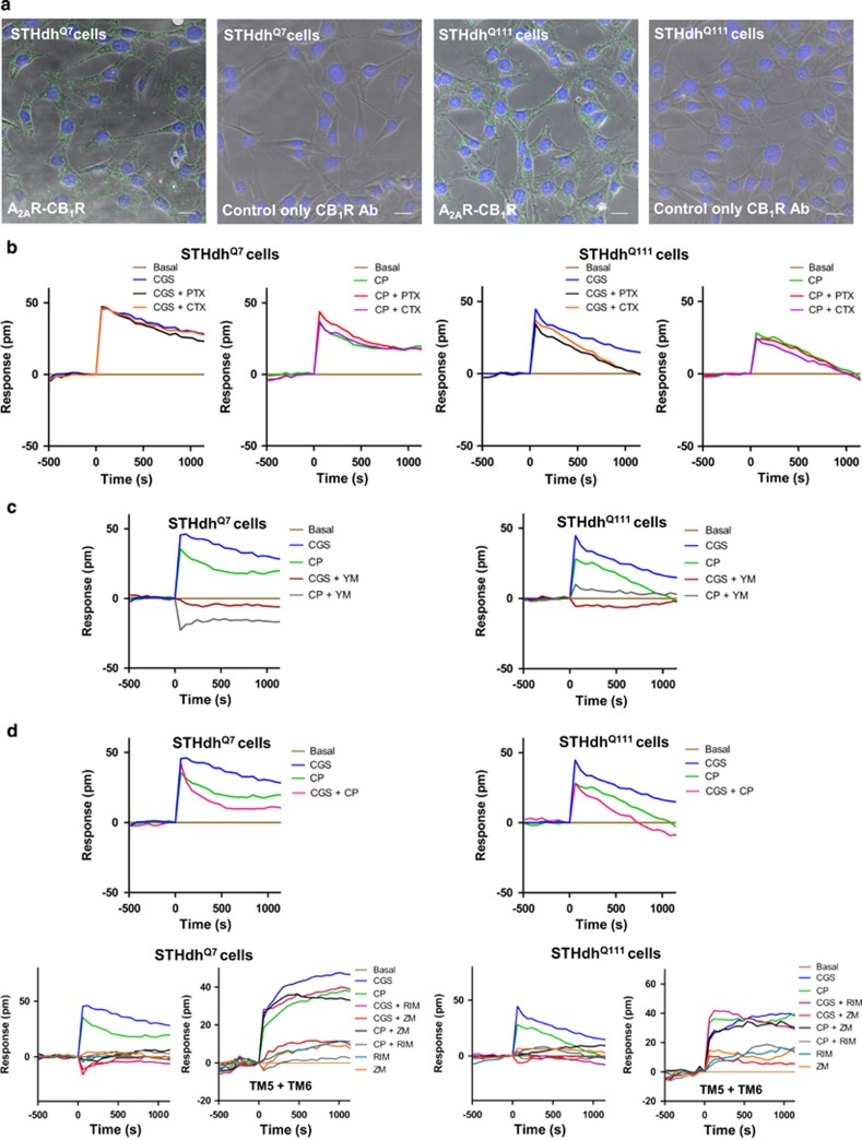
A2AR-CB1R heteromers expressed in wild-type STHdhQ7 and mutant huntingtin-expressing STHdhQ111 striatal neuroblasts signal via Gq protein rather than Gi or Gs protein. (a) PLA assays were performed in STHdhQ7 and STHdhQ111 cells. A2AR-CB1R heteromers are shown as green dots. Nuclei are colored in blue by DAPI staining. Controls in the absence of anti-A2AR primary antibody were also performed. Representative pictures are shown. Scale bar: 20 μm. (b) Dynamic mass redistribution (DMR) assays were performed in STHdhQ7 and STHdhQ111 cells pretreated overnight with vehicle, pertussis toxin (PTX; 10 ng/ml) or cholera toxin (CTX; 100 ng/ml), and further treated with vehicle, the A2AR agonist CGS21680 (1 μM) or the CB1R agonist CP-55,940 (1 μM). (c) DMR assays in STHdhQ7 and STHdhQ111 cells preincubated for 30 min with vehicle or the Gq protein inhibitor YM-254890 (1 μM), and then activated with the A2AR agonist CGS21680 (1 μM) or the CB1R agonist CP-55,940 (1 μM). (d) DMR assays showing negative cross-talk (top panels) and cross-antagonism (bottom panels) between A2AR and CB1R signaling. STHdhQ7 and STHdhQ111 cells were not pre-treated (top panels) or pre-treated for 4 h with medium (left bottom panels) or with 4 μM TM5 plus TM6 (right bottom panels) before incubation for 30 min with vehicle, the CB1R antagonist SR141716 (RIM; 1 μM) or the A2AR antagonist ZM241385 (1 μM), and then activated with vehicle, CGS21680 (1 μM) or CP-55,940 (1 μM). (b–d) The resulting shifts of reflected light wavelength (pm) were monitored over time. Each panel is a representative experiment of n=3 different experiments. Each curve is the mean of a representative optical trace experiment carried out in triplicates.

