Figure 1.
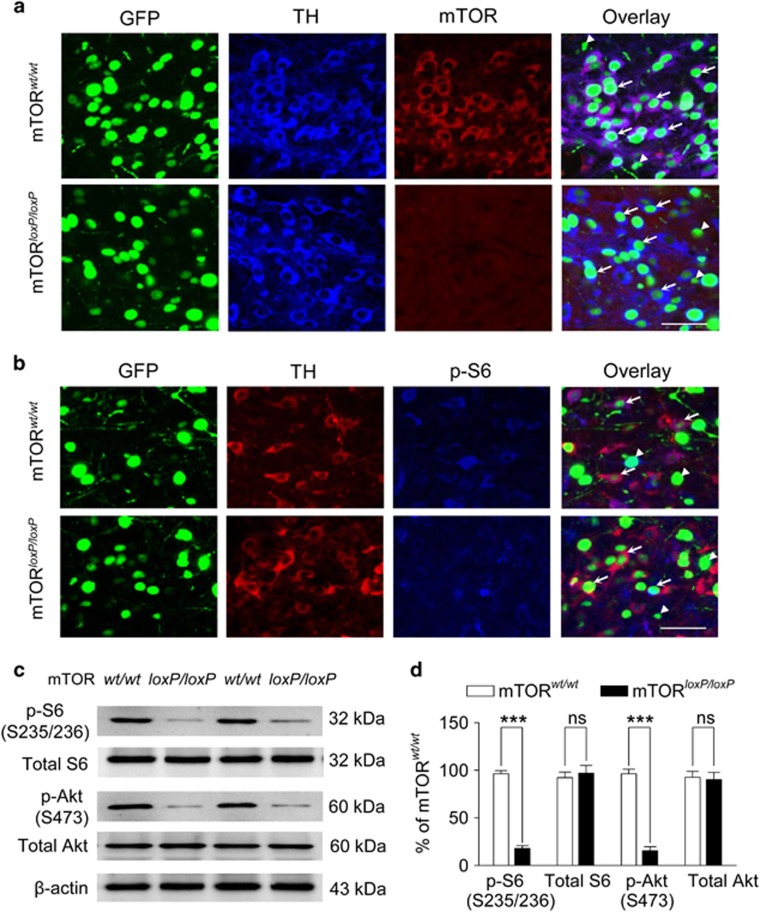
AAV2-Cre-GFP-mediated deletion of mTOR in the VTA. VTA immunostaining following mTOR deletion. (a) Immunofluorescence labeling indicates that mTOR expression remains in mTORwt/wt mice (n=3) but not mTORloxP/loxP mice (n=4) 2–3 weeks after intra-VTA injection of AAV2-Cre-GFP. Scale bar, 50 μm. Arrows: representative dopamine neurons; arrowheads, representative non-dopamine neurons. (b) Immunofluorescence labeling indicates that mTOR deletion decreased phosphorylated S6 kinase (p-S6 S235/236) expression in the VTA (n=2 mice). (c, d) Western blots (c) and normalized data (d) revealed that mTOR deletion led to decreases in p-S6 S235/236 (mTORC1-dependent) and p-Akt S473 (mTORC2-dependent) in the VTA (n=5 mice/group; ***P<0.001, ns, not significant).

