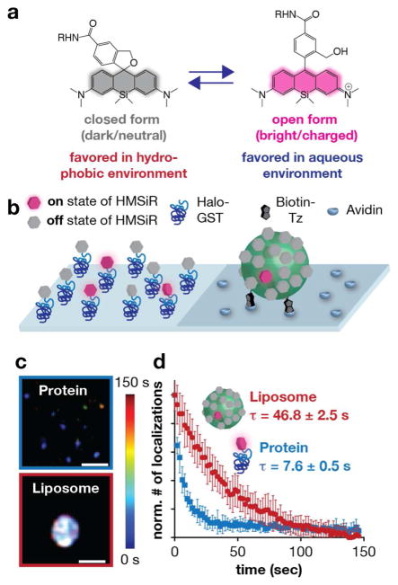
Figure 5.
In vitro evaluation of HMSiR in a membrane environment. a) HMSiR equilibrates between a closed, cyclized form that dark (OFF) and an open (ON)form that is bright. Like SiR-CO2H, the position of the HMSiR equilibrium depends on pH and hydrophobicity. b) Cartoon of an in vitro experiment designed to assess whether the HMSiR ON/OFF fraction is affected by hydrophobicity. HMSiR was immobilized to a glass surface via either a protein (generated upon reaction of HMSiR-CA and Halo-GST) or a liposome (generated upon reaction of Cer-TCO with HMSiR-Tz). c) Super-resolution images of HMSiR immobilized as described in b). Rainbow colored temporal look-up table. Scale bar: 200 nm. d) Plot illustrating the normalized number of localizations observed as a function of time when HMSiR was immobilized to a protein, within an aqueous environment, and within a liposome. τ values were calculated from a single exponential fit (mean ± SEM). Figure adapted with permissionfrom Takakura et al.40

