Figure 4.
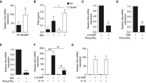
MyD88 signaling negatively regulates type I IFN expression in B cells. (A) Ifnb mRNA expression in B cells purified from the spleen of Myd88−/− (open bars) and wild type (WT, solid bars) control mice on stimulation for 24 hours with Mycobacterium tuberculosis (Mtb) (multiplicity of infection = 0.3); fold change represents expression after stimulation relative to respective expression before stimulation (set to 1) (n = 10 mice per group). (B) Concentration of IFNβ in the supernatants of naive splenic B cells from WT or Myd88−/− mice stimulated for 6 days or not with Mtb (n = 3). (C–E) Ifnb mRNA expression in B cells purified from the spleen of WT mice (n = 4–8 mice per group) on 24-hour in vitro stimulation with either c-di-AMP (C), TDB (D) or Mtb (E) in the presence (+) or absence (−) of the TLR2 agonist Pam3CSK4. (F) B cells from WT mice were stimulated with c-di-AMP and/or IL1β, then IFNβ expression was analyzed at the mRNA level (n = 6). (G) As in F, except that Myd88−/− B cells were used (n = 4). Data represent mean ± SEM and were analyzed by using the two-tailed Mann-Whitney test or the two-tailed Wilcoxon test (B–E) (*P ≤ 0.05; **P ≤ 0.01; ***P ≤ 0.001). The two-tailed Student paired t test was used for A and F (#P ≤ 0.05; ##P ≤ 0.01; ###P ≤ 0.001). TDB = trehalose-6,6-dibehenate.

