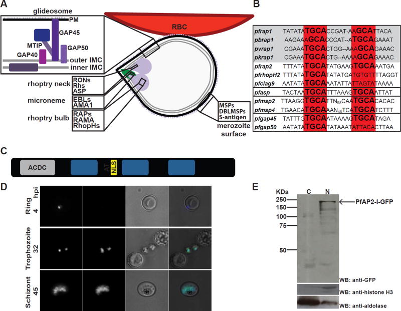
Figure 1. PfAP2-I is a nuclear protein that may bind a conserved TGCA DNA motif upstream of some invasion genes.
A- Merozoite attached to the RBC surface with the micronemes, rhoptries, merozoite surface, plasma membrane (PM), inner and outer inner membrane complex (IMC) depicted. Proteins found in the glideosome, merozoite surface, microneme, rhoptry neck and rhoptry bulb are highlighted. B- The NGGTGCA DNA sequence motif is conserved in invasion-related gene promoters across P. falciparum (Pf), P. berghei (Pb), P. vivax (Pv) or P. knowlesi (Pk) (grey box). The four black boxes (from top to bottom) contain rhoptry promoters, the pfasp rhoptry neck promoter, merozoite surface (msp) promoters, and glideosome (gap) promoters (adapted from (Young et al., 2008)) (see also figure S1A). C- The PfAP2-I protein structure contains a putative ACDC domain, the three AP2 DNA-binding domains, an AT-hook and a nuclear localization signal (NLS) (see also figure S2). D- Live fluorescence microscopy of synchronized parasites shows that PfAP2-I-GFP (see figure S1B) localizes to the nucleus of trophozoite and schizont stage parasites but is not detected in ring stages. Hoechst was used as a nuclear marker. BF denotes bright field. E- Nuclear fractionation followed by Western blot of schizont-stage PfAP2-I-GFP parasites confirms nuclear localization of PfAP2-I-GFP. Anti-histone H3 and anti-aldolase were used as nuclear and cytosolic markers, respectively. C: cytosol, N: nucleus.

