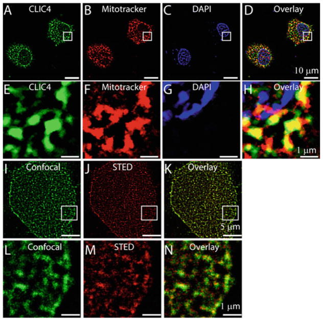
Fig. 2.
Localization of CLIC4 in H9C2 cells. H9C2 cells loaded with mitotracker (b) were labeled with anti-CLIC4 antibody (a), and stained with DAPI (c). d is merge image of a, b, and c showing colocalization of CLIC4 to the mitochondria of H9C2 cells. e, f, g, and h are enlarged images of the squared regions in a, b, c, and d, respectively. STED microscopy of H9C2 cells showing expression of CLIC4 in the mitochondria (j, k). Confocal image of H9C2 cells labeled with anti-CLIC4 antibody (i). Corresponding STED image (j), overlay of i and j highlights increased resolution of CLIC4 localization to mitochondria with STED microscopy (k). l, m, and n are enlarged images of the squared region in i, j, and k, respectively

