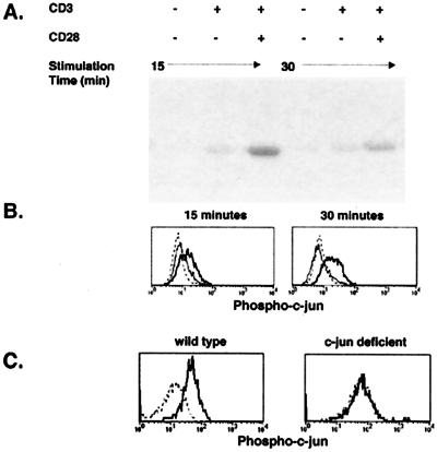
Figure 1.
Comparison of phospho-c-jun detection by flow cytometry and JNK enzymatic activity in naive T cells stimulated in vitro with mAbs. Purified DO11.10 T cells were stimulated for 15 or 30 min with control hamster IgG, anti-CD3, or anti-CD3 and anti-CD28 mAbs. (A) Whole-cell lysates from stimulated cells were incubated with GST-c-jun beads and analyzed for JNK activity. (B) Intracellular staining of phosphorylated c-jun protein in cells stimulated with control IgG (dashed line), anti-CD3 (thin line), or anti-CD3 and anti-CD28 mAbs (bold line). (C) Intracellular staining of phosphorylated c-jun protein in normal or c-jun-deficient mouse embryo fibroblasts stimulated for 10 min with EGF (bold line) or nothing (dashed line).

