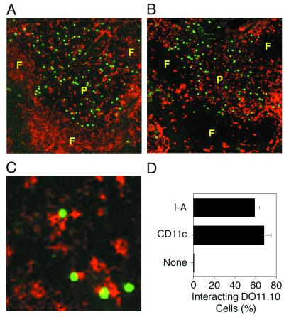
Figure 3.
Naive CD4 T cells are located near I-Ad-expressing cells in vivo. The locations of DO11.10 CD4 T cells (shown green) and cells expressing anti-I-Ad (shown red in A) or anti-CD11c (shown red in B) in thin sections of the spleen are shown. A and B show adjacent sections containing the same white pulp chord with a perimeter of B-cell-rich follicles (F) surrounding a central periarteriolar lymphoid sheath (P). A magnified view of a periarteriolar lymphoid sheath is shown in C with DO11.10 T cells shown green and I-Ad-expressing cells shown red. The areas of yellow color indicate significant overlap between the two cell types. D shows the mean percentage of DO11.10 T cells (± the standard deviation) found interacting with cells stained with I-Ad, CD11c, or control Ig, as evidenced by the presence of yellow color. At least 200 cells from two to four white pulp chords were counted in each group.

