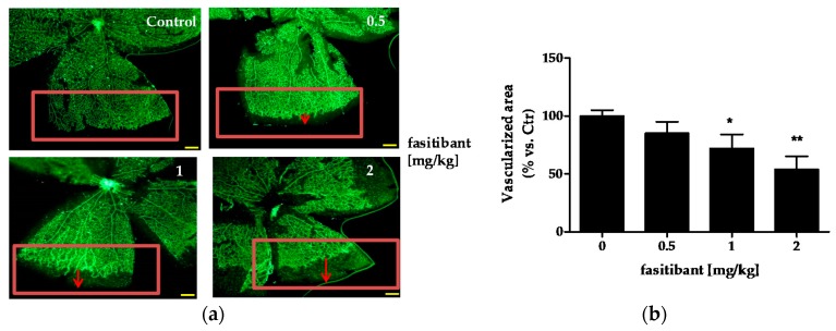
Figure 1.
Fasitibant reduces the extent of retinal vascularization in mouse pups. (a) pups were treated with or without fasitibant (NaCl 0.9% = Control; fasitibant, 0.5 to 2 mg/kg, in 50 µL, i.p.) and then retinas were dissected at P8 and stained with isolectin, IB4-488. Magnitude 4×, scale bar = 100 µm; (b) quantification of retinal vessels. Retinal whole-mounts from P8 pups were stained for endothelial cells with IB4-488. For measurement of the vascularized area at the retinal edges, the retinal edges were outlined with image-processing software (Photoshop Adobe Systems, Adobe Photoshop Elements 11). The density of vascularization in the outlined areas was quantified by ImageJ software, version 2.0.0-rc-43/1.50e, U.S. National Institutes of Health, Bethesda, MD, USA; and expressed in relation to the control areas (% vs. Ctr). Data are the mean of 10 outlined areas, obtained from at least five flower-like structures. Arrows indicate the distance from vascularized area to retinal edge. * p < 0.05, ** p < 0.01 vs. Ctr.

