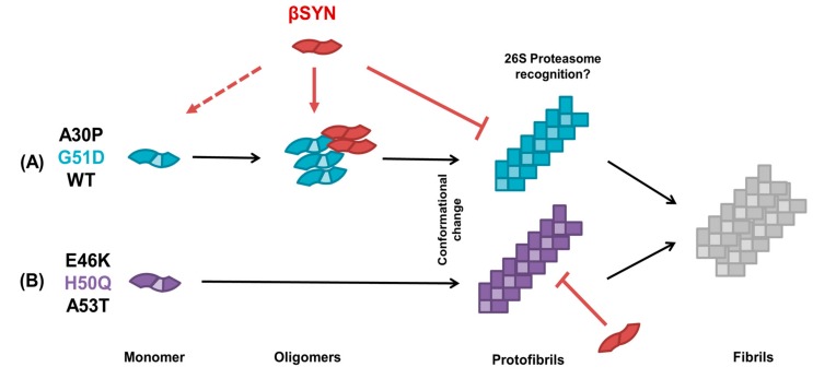
Figure 6.
Proposed model of modulation of α-synuclein aggregation by its putative “natural negative regulator” β-synuclein. The model shows two distinct pathways of aggregation for the different αS mutants, in (A) and (B). αS monomers are depicted as blue and purple coloured blocks with a light-coloured core representing the NAC domain, which is absent in βS, represented by red blocks. Arrows represent the proposed model’s sequence; dashed lines represent discussion points in this model for future studies; capped ends depict no interaction. Monomeric and oligomeric forms are represented unfolded, and a conformational change is required for α-synuclein to display pre-fibrillar and fibrillar species.

