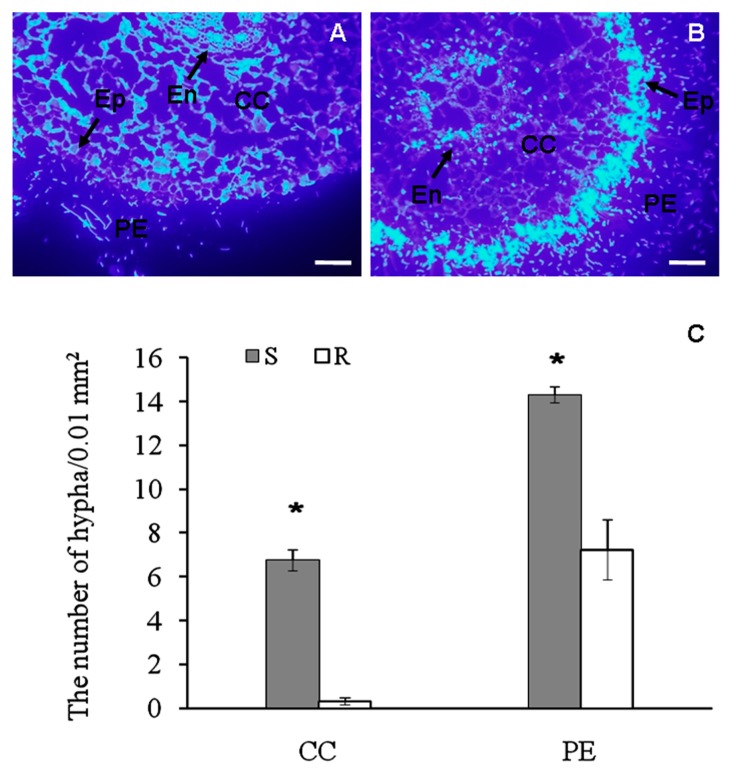
Figure 1.
The tissue-specific pathogen spreading in roots of infected banana (Musa spp. AAA) seedlings. (A,B) Visualization of Fusarium oxysporum f. sp. cubense hyphae by calcofluor white stain in the resistant cultivar “Yueyoukang 1” (A) and susceptible “Baxijiao” (B) 48 h after infection; (C) Quantification of hyphae in A and B. Bars represent 100 μm. CC, cortical cells; Ep, epidermis; En, endodermis; PE, periphery of epidermis; R, resistant cultivar “Yueyoukang 1”; S, susceptible cultivar “Baxijiao”. Quantitative data represent an average of three biological replicates. Comparison of groups was conducted using a paired t-test of variance. Values marked with * were considered significant at p < 0.05.

