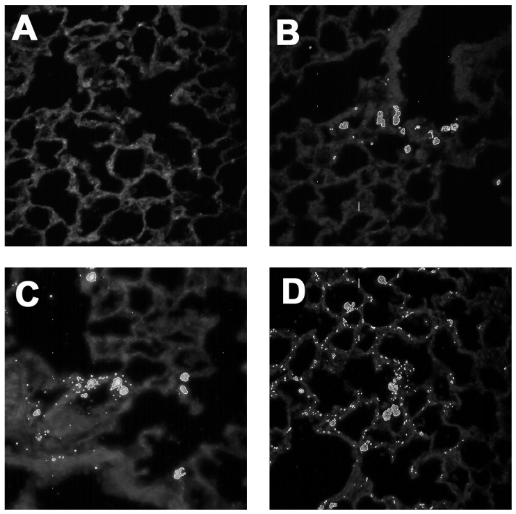
Figure 5.
CytoViva dark-field images (400×) using Particle Fitting Function to locate MWCNT in lung tissue following a three-day particle exposure: (A) no particle dispersion media control; (B) source MWCNTs isolated mainly in alveolar macrophages. (C) carboxylated MWCNT20.6 (20 min) starting to show some distribution in the lung lining in addition to AM cell collection; and (D) maximally carboxylated MWCNT14.7 (120 min) demonstrates particle dispersion throughout the lung lining indicating epithelial cell involvement.

